Chlorine »
PDB 2r7k-2rh8 »
2r8x »
Chlorine in PDB 2r8x: Crystal Structure of Yrbi Phosphatase From Escherichia Coli
Enzymatic activity of Crystal Structure of Yrbi Phosphatase From Escherichia Coli
All present enzymatic activity of Crystal Structure of Yrbi Phosphatase From Escherichia Coli:
3.1.3.45;
3.1.3.45;
Protein crystallography data
The structure of Crystal Structure of Yrbi Phosphatase From Escherichia Coli, PDB code: 2r8x
was solved by
O.V.Tsodikov,
P.Aggarwal,
J.R.Rubin,
J.A.Stuckey,
R.W.Woodard,
T.Biswas,
with X-Ray Crystallography technique. A brief refinement statistics is given in the table below:
| Resolution Low / High (Å) | 30.00 / 2.60 |
| Space group | P 1 21 1 |
| Cell size a, b, c (Å), α, β, γ (°) | 85.071, 156.959, 114.407, 90.00, 96.54, 90.00 |
| R / Rfree (%) | 20.1 / 23.6 |
Chlorine Binding Sites:
The binding sites of Chlorine atom in the Crystal Structure of Yrbi Phosphatase From Escherichia Coli
(pdb code 2r8x). This binding sites where shown within
5.0 Angstroms radius around Chlorine atom.
In total 4 binding sites of Chlorine where determined in the Crystal Structure of Yrbi Phosphatase From Escherichia Coli, PDB code: 2r8x:
Jump to Chlorine binding site number: 1; 2; 3; 4;
In total 4 binding sites of Chlorine where determined in the Crystal Structure of Yrbi Phosphatase From Escherichia Coli, PDB code: 2r8x:
Jump to Chlorine binding site number: 1; 2; 3; 4;
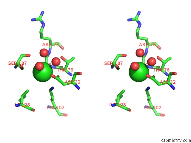
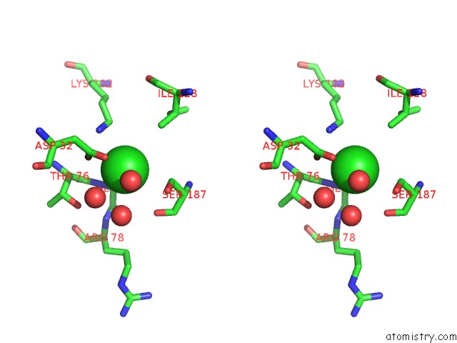
Chlorine binding site 1 out of 4 in 2r8x
Go back to
Chlorine binding site 1 out
of 4 in the Crystal Structure of Yrbi Phosphatase From Escherichia Coli

Mono view
Stereo pair view
Mono view
Stereo pair view
A full contact list of Chlorine with other atoms in the Cl binding
site number 1 of Crystal Structure of Yrbi Phosphatase From Escherichia Coli within 5.0Å range:
|
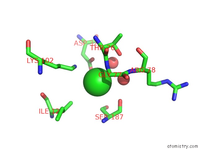
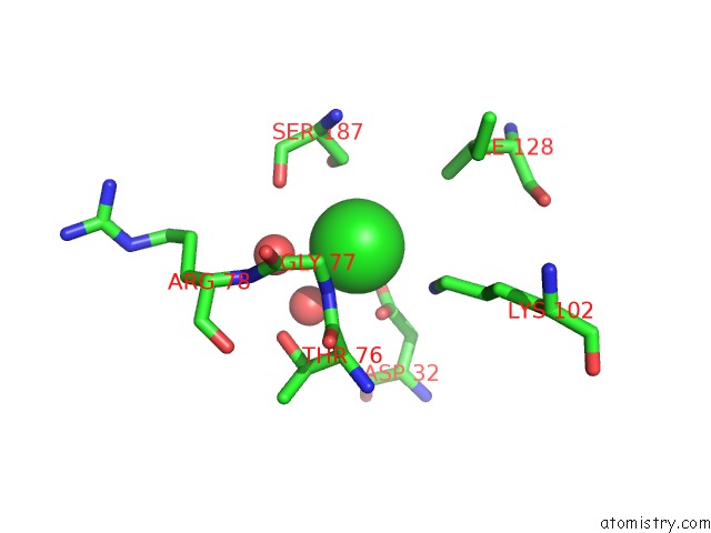
Chlorine binding site 2 out of 4 in 2r8x
Go back to
Chlorine binding site 2 out
of 4 in the Crystal Structure of Yrbi Phosphatase From Escherichia Coli
Mono view
Stereo pair view
Mono view
Stereo pair view
A full contact list of Chlorine with other atoms in the Cl binding
site number 2 of Crystal Structure of Yrbi Phosphatase From Escherichia Coli within 5.0Å range:
|
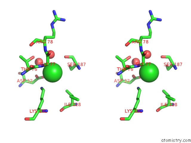
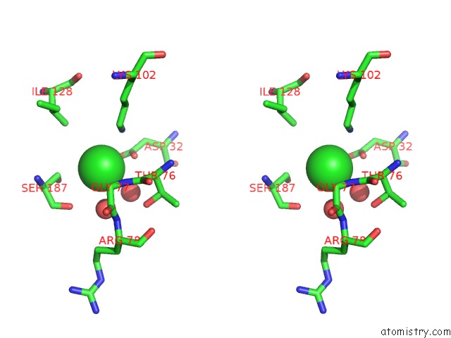
Chlorine binding site 3 out of 4 in 2r8x
Go back to
Chlorine binding site 3 out
of 4 in the Crystal Structure of Yrbi Phosphatase From Escherichia Coli
Mono view
Stereo pair view
Mono view
Stereo pair view
A full contact list of Chlorine with other atoms in the Cl binding
site number 3 of Crystal Structure of Yrbi Phosphatase From Escherichia Coli within 5.0Å range:
|
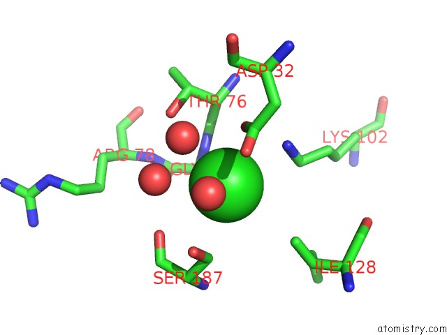
Chlorine binding site 4 out of 4 in 2r8x
Go back to
Chlorine binding site 4 out
of 4 in the Crystal Structure of Yrbi Phosphatase From Escherichia Coli
Mono view
Stereo pair view
Mono view
Stereo pair view
A full contact list of Chlorine with other atoms in the Cl binding
site number 4 of Crystal Structure of Yrbi Phosphatase From Escherichia Coli within 5.0Å range:
|
Reference:
T.Biswas,
L.Yi,
P.Aggarwal,
J.Wu,
J.R.Rubin,
J.A.Stuckey,
R.W.Woodard,
O.V.Tsodikov.
The Tail of Kdsc: Conformational Changes Control the Activity of A Haloacid Dehalogenase Superfamily Phosphatase. J.Biol.Chem. V. 284 30594 2009.
ISSN: ISSN 0021-9258
PubMed: 19726684
DOI: 10.1074/JBC.M109.012278
Page generated: Sat Jul 20 11:09:26 2024
ISSN: ISSN 0021-9258
PubMed: 19726684
DOI: 10.1074/JBC.M109.012278
Last articles
Zn in 9J0NZn in 9J0O
Zn in 9J0P
Zn in 9FJX
Zn in 9EKB
Zn in 9C0F
Zn in 9CAH
Zn in 9CH0
Zn in 9CH3
Zn in 9CH1