Chlorine »
PDB 2r7k-2rh8 »
2rg8 »
Chlorine in PDB 2rg8: Crystal Structure of Programmed For Cell Death 4 Middle MA3 Domain
Protein crystallography data
The structure of Crystal Structure of Programmed For Cell Death 4 Middle MA3 Domain, PDB code: 2rg8
was solved by
R.Garces,
C.Suzuki,
G.Wagner,
with X-Ray Crystallography technique. A brief refinement statistics is given in the table below:
| Resolution Low / High (Å) | 35.68 / 1.80 |
| Space group | P 21 21 21 |
| Cell size a, b, c (Å), α, β, γ (°) | 37.680, 70.060, 110.880, 90.00, 90.00, 90.00 |
| R / Rfree (%) | 20.2 / 25.1 |
Other elements in 2rg8:
The structure of Crystal Structure of Programmed For Cell Death 4 Middle MA3 Domain also contains other interesting chemical elements:
| Sodium | (Na) | 3 atoms |
Chlorine Binding Sites:
The binding sites of Chlorine atom in the Crystal Structure of Programmed For Cell Death 4 Middle MA3 Domain
(pdb code 2rg8). This binding sites where shown within
5.0 Angstroms radius around Chlorine atom.
In total 2 binding sites of Chlorine where determined in the Crystal Structure of Programmed For Cell Death 4 Middle MA3 Domain, PDB code: 2rg8:
Jump to Chlorine binding site number: 1; 2;
In total 2 binding sites of Chlorine where determined in the Crystal Structure of Programmed For Cell Death 4 Middle MA3 Domain, PDB code: 2rg8:
Jump to Chlorine binding site number: 1; 2;
Chlorine binding site 1 out of 2 in 2rg8
Go back to
Chlorine binding site 1 out
of 2 in the Crystal Structure of Programmed For Cell Death 4 Middle MA3 Domain
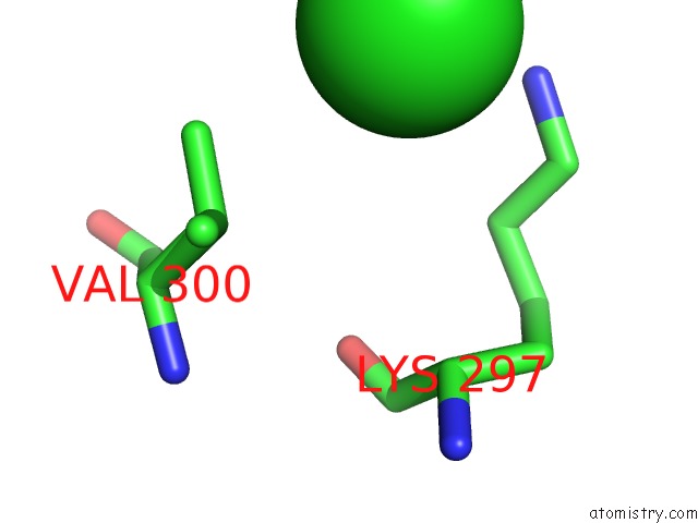
Mono view
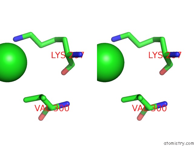
Stereo pair view
Mono view
Stereo pair view
A full contact list of Chlorine with other atoms in the Cl binding
site number 1 of Crystal Structure of Programmed For Cell Death 4 Middle MA3 Domain within 5.0Å range:
|
Chlorine binding site 2 out of 2 in 2rg8
Go back to
Chlorine binding site 2 out
of 2 in the Crystal Structure of Programmed For Cell Death 4 Middle MA3 Domain
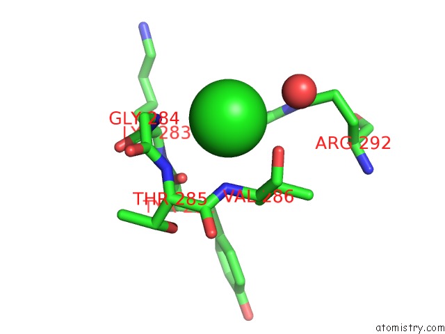
Mono view
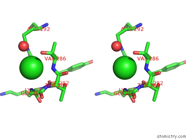
Stereo pair view
Mono view
Stereo pair view
A full contact list of Chlorine with other atoms in the Cl binding
site number 2 of Crystal Structure of Programmed For Cell Death 4 Middle MA3 Domain within 5.0Å range:
|
Reference:
C.Suzuki,
R.G.Garces,
K.A.Edmonds,
S.Hiller,
S.G.Hyberts,
A.Marintchev,
G.Wagner.
PDCD4 Inhibits Translation Initiation By Binding to EIF4A Using Both Its MA3 Domains. Proc.Natl.Acad.Sci.Usa V. 105 3274 2008.
ISSN: ISSN 0027-8424
PubMed: 18296639
DOI: 10.1073/PNAS.0712235105
Page generated: Sat Jul 20 11:18:48 2024
ISSN: ISSN 0027-8424
PubMed: 18296639
DOI: 10.1073/PNAS.0712235105
Last articles
Zn in 9J0NZn in 9J0O
Zn in 9J0P
Zn in 9FJX
Zn in 9EKB
Zn in 9C0F
Zn in 9CAH
Zn in 9CH0
Zn in 9CH3
Zn in 9CH1