Chlorine »
PDB 2rha-2uxn »
2uw7 »
Chlorine in PDB 2uw7: Structure of Pka-Pkb Chimera Complexed with 4-(4-Chloro- Phenyl)-4-(4-(1H-Pyrazol-4-Yl)-Phenyl)-Piperidine
Protein crystallography data
The structure of Structure of Pka-Pkb Chimera Complexed with 4-(4-Chloro- Phenyl)-4-(4-(1H-Pyrazol-4-Yl)-Phenyl)-Piperidine, PDB code: 2uw7
was solved by
T.G.Davies,
G.Saxty,
S.J.Woodhead,
V.Berdini,
M.L.Verdonk,
P.G.Wyatt,
R.G.Boyle,
D.Barford,
R.Downham,
M.D.Garrett,
R.A.Carr,
with X-Ray Crystallography technique. A brief refinement statistics is given in the table below:
| Resolution Low / High (Å) | 27.43 / 2.1 |
| Space group | P 21 21 21 |
| Cell size a, b, c (Å), α, β, γ (°) | 72.940, 75.190, 80.205, 90.00, 90.00, 90.00 |
| R / Rfree (%) | 23.5 / 31.8 |
Chlorine Binding Sites:
The binding sites of Chlorine atom in the Structure of Pka-Pkb Chimera Complexed with 4-(4-Chloro- Phenyl)-4-(4-(1H-Pyrazol-4-Yl)-Phenyl)-Piperidine
(pdb code 2uw7). This binding sites where shown within
5.0 Angstroms radius around Chlorine atom.
In total only one binding site of Chlorine was determined in the Structure of Pka-Pkb Chimera Complexed with 4-(4-Chloro- Phenyl)-4-(4-(1H-Pyrazol-4-Yl)-Phenyl)-Piperidine, PDB code: 2uw7:
In total only one binding site of Chlorine was determined in the Structure of Pka-Pkb Chimera Complexed with 4-(4-Chloro- Phenyl)-4-(4-(1H-Pyrazol-4-Yl)-Phenyl)-Piperidine, PDB code: 2uw7:
Chlorine binding site 1 out of 1 in 2uw7
Go back to
Chlorine binding site 1 out
of 1 in the Structure of Pka-Pkb Chimera Complexed with 4-(4-Chloro- Phenyl)-4-(4-(1H-Pyrazol-4-Yl)-Phenyl)-Piperidine
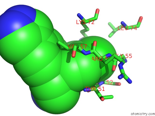
Mono view
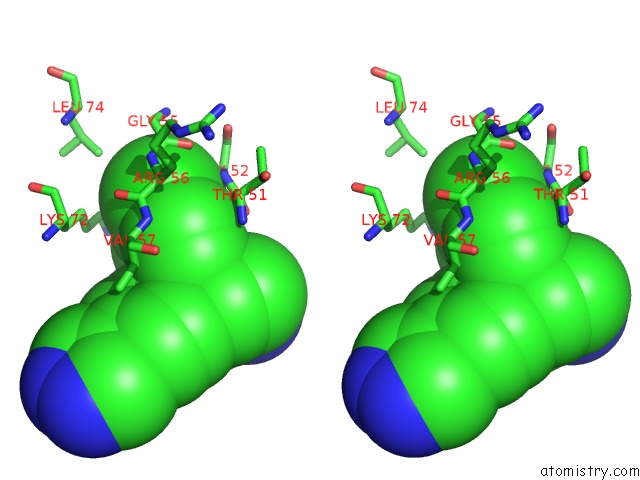
Stereo pair view
Mono view
Stereo pair view
A full contact list of Chlorine with other atoms in the Cl binding
site number 1 of Structure of Pka-Pkb Chimera Complexed with 4-(4-Chloro- Phenyl)-4-(4-(1H-Pyrazol-4-Yl)-Phenyl)-Piperidine within 5.0Å range:
|
Reference:
G.Saxty,
S.J.Woodhead,
V.Berdini,
T.G.Davies,
M.L.Verdonk,
P.G.Wyatt,
R.G.Boyle,
D.Barford,
R.Downham,
M.D.Garrett,
R.A.Carr.
Identification of Inhibitors of Protein Kinase B Using Fragment-Based Lead Discovery J.Med.Chem. V. 50 2293 2007.
ISSN: ISSN 0022-2623
PubMed: 17451234
DOI: 10.1021/JM070091B
Page generated: Sat Jul 20 11:25:38 2024
ISSN: ISSN 0022-2623
PubMed: 17451234
DOI: 10.1021/JM070091B
Last articles
Zn in 9J0NZn in 9J0O
Zn in 9J0P
Zn in 9FJX
Zn in 9EKB
Zn in 9C0F
Zn in 9CAH
Zn in 9CH0
Zn in 9CH3
Zn in 9CH1