Chlorine »
PDB 2uxp-2v79 »
2v0d »
Chlorine in PDB 2v0d: Crystal Structure of Human CDK2 Complexed with A Thiazolidinone Inhibitor
Enzymatic activity of Crystal Structure of Human CDK2 Complexed with A Thiazolidinone Inhibitor
All present enzymatic activity of Crystal Structure of Human CDK2 Complexed with A Thiazolidinone Inhibitor:
2.7.1.37; 2.7.11.22;
2.7.1.37; 2.7.11.22;
Protein crystallography data
The structure of Crystal Structure of Human CDK2 Complexed with A Thiazolidinone Inhibitor, PDB code: 2v0d
was solved by
C.M.Richardson,
P.Dokurno,
J.B.Murray,
A.E.Surgenor,
with X-Ray Crystallography technique. A brief refinement statistics is given in the table below:
| Resolution Low / High (Å) | 8.00 / 2.20 |
| Space group | P 21 21 21 |
| Cell size a, b, c (Å), α, β, γ (°) | 53.459, 71.850, 72.011, 90.00, 90.00, 90.00 |
| R / Rfree (%) | 19.3 / 28.6 |
Chlorine Binding Sites:
The binding sites of Chlorine atom in the Crystal Structure of Human CDK2 Complexed with A Thiazolidinone Inhibitor
(pdb code 2v0d). This binding sites where shown within
5.0 Angstroms radius around Chlorine atom.
In total only one binding site of Chlorine was determined in the Crystal Structure of Human CDK2 Complexed with A Thiazolidinone Inhibitor, PDB code: 2v0d:
In total only one binding site of Chlorine was determined in the Crystal Structure of Human CDK2 Complexed with A Thiazolidinone Inhibitor, PDB code: 2v0d:
Chlorine binding site 1 out of 1 in 2v0d
Go back to
Chlorine binding site 1 out
of 1 in the Crystal Structure of Human CDK2 Complexed with A Thiazolidinone Inhibitor
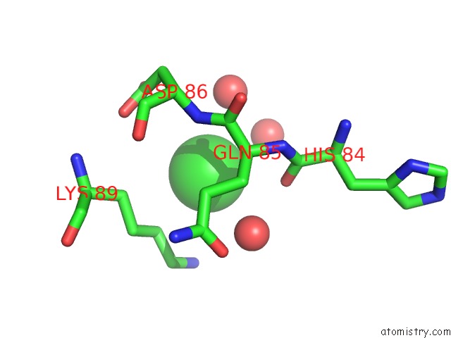
Mono view
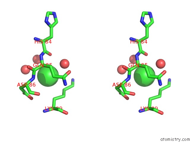
Stereo pair view
Mono view
Stereo pair view
A full contact list of Chlorine with other atoms in the Cl binding
site number 1 of Crystal Structure of Human CDK2 Complexed with A Thiazolidinone Inhibitor within 5.0Å range:
|
Reference:
C.M.Richardson,
C.L.Nunns,
D.S.Williamson,
M.J.Parratt,
P.Dokurno,
R.Howes,
J.Borgognoni,
M.J.Drysdale,
H.Finch,
R.E.Hubbard,
P.S.Jackson,
P.Kierstan,
G.Lentzen,
J.D.Moore,
J.B.Murray,
H.Simmonite,
A.E.Surgenor,
C.J.Torrance.
Discovery of A Potent CDK2 Inhibitor with A Novel Binding Mode, Using Virtual Screening and Initial, Structure-Guided Lead Scoping. Bioorg.Med.Chem.Lett. V. 17 3880 2007.
ISSN: ISSN 0960-894X
PubMed: 17570665
DOI: 10.1016/J.BMCL.2007.04.110
Page generated: Sat Jul 20 11:30:02 2024
ISSN: ISSN 0960-894X
PubMed: 17570665
DOI: 10.1016/J.BMCL.2007.04.110
Last articles
Zn in 9MJ5Zn in 9HNW
Zn in 9G0L
Zn in 9FNE
Zn in 9DZN
Zn in 9E0I
Zn in 9D32
Zn in 9DAK
Zn in 8ZXC
Zn in 8ZUF