Chlorine »
PDB 2uxp-2v79 »
2v3p »
Chlorine in PDB 2v3p: Crystallographic Analysis of Beta-Axial Ligand Substitutions in Cobalamin Bound to Transcobalamin
Protein crystallography data
The structure of Crystallographic Analysis of Beta-Axial Ligand Substitutions in Cobalamin Bound to Transcobalamin, PDB code: 2v3p
was solved by
J.Wuerges,
S.Geremia,
L.Randaccio,
with X-Ray Crystallography technique. A brief refinement statistics is given in the table below:
| Resolution Low / High (Å) | 28.98 / 2.90 |
| Space group | P 31 2 1 |
| Cell size a, b, c (Å), α, β, γ (°) | 100.390, 100.390, 129.742, 90.00, 90.00, 120.00 |
| R / Rfree (%) | 19.4 / 25 |
Other elements in 2v3p:
The structure of Crystallographic Analysis of Beta-Axial Ligand Substitutions in Cobalamin Bound to Transcobalamin also contains other interesting chemical elements:
| Cobalt | (Co) | 1 atom |
Chlorine Binding Sites:
The binding sites of Chlorine atom in the Crystallographic Analysis of Beta-Axial Ligand Substitutions in Cobalamin Bound to Transcobalamin
(pdb code 2v3p). This binding sites where shown within
5.0 Angstroms radius around Chlorine atom.
In total 3 binding sites of Chlorine where determined in the Crystallographic Analysis of Beta-Axial Ligand Substitutions in Cobalamin Bound to Transcobalamin, PDB code: 2v3p:
Jump to Chlorine binding site number: 1; 2; 3;
In total 3 binding sites of Chlorine where determined in the Crystallographic Analysis of Beta-Axial Ligand Substitutions in Cobalamin Bound to Transcobalamin, PDB code: 2v3p:
Jump to Chlorine binding site number: 1; 2; 3;
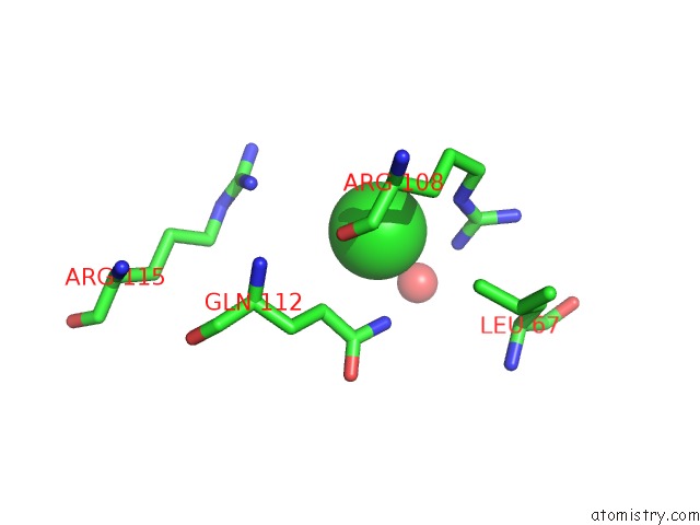
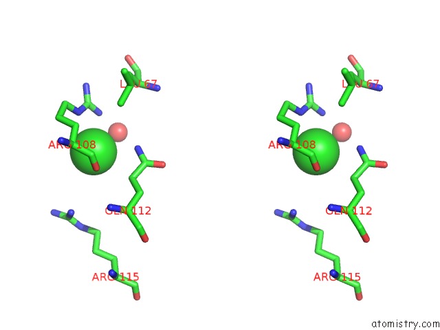
Chlorine binding site 1 out of 3 in 2v3p
Go back to
Chlorine binding site 1 out
of 3 in the Crystallographic Analysis of Beta-Axial Ligand Substitutions in Cobalamin Bound to Transcobalamin
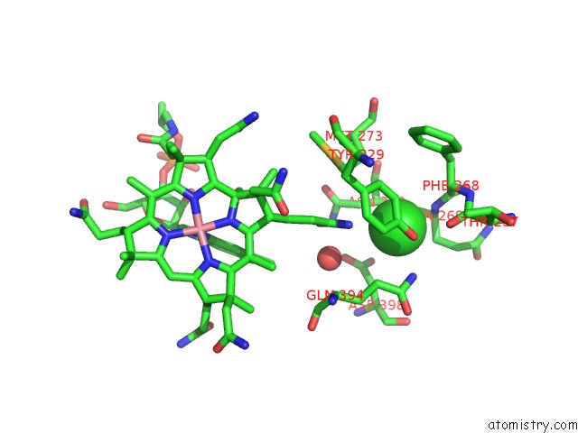
Mono view
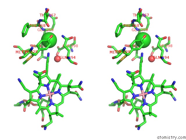
Stereo pair view
Mono view
Stereo pair view
A full contact list of Chlorine with other atoms in the Cl binding
site number 1 of Crystallographic Analysis of Beta-Axial Ligand Substitutions in Cobalamin Bound to Transcobalamin within 5.0Å range:
|
Chlorine binding site 2 out of 3 in 2v3p
Go back to
Chlorine binding site 2 out
of 3 in the Crystallographic Analysis of Beta-Axial Ligand Substitutions in Cobalamin Bound to Transcobalamin
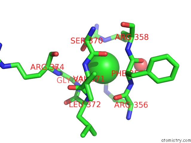
Mono view
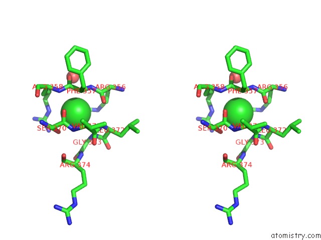
Stereo pair view
Mono view
Stereo pair view
A full contact list of Chlorine with other atoms in the Cl binding
site number 2 of Crystallographic Analysis of Beta-Axial Ligand Substitutions in Cobalamin Bound to Transcobalamin within 5.0Å range:
|
Chlorine binding site 3 out of 3 in 2v3p
Go back to
Chlorine binding site 3 out
of 3 in the Crystallographic Analysis of Beta-Axial Ligand Substitutions in Cobalamin Bound to Transcobalamin
Mono view
Stereo pair view
Mono view
Stereo pair view
A full contact list of Chlorine with other atoms in the Cl binding
site number 3 of Crystallographic Analysis of Beta-Axial Ligand Substitutions in Cobalamin Bound to Transcobalamin within 5.0Å range:
|
Reference:
J.Wuerges,
S.Geremia,
S.N.Fedosov,
L.Randaccio.
Vitamin B12 Transport Proteins: Crystallographic Analysis of Beta-Axial Ligand Substitutions in Cobalamin Bound to Transcobalamin. Iubmb Life V. 59 722 2007.
ISSN: ISSN 1521-6543
PubMed: 17943552
DOI: 10.1080/15216540701673413
Page generated: Sat Jul 20 11:35:05 2024
ISSN: ISSN 1521-6543
PubMed: 17943552
DOI: 10.1080/15216540701673413
Last articles
Zn in 9MJ5Zn in 9HNW
Zn in 9G0L
Zn in 9FNE
Zn in 9DZN
Zn in 9E0I
Zn in 9D32
Zn in 9DAK
Zn in 8ZXC
Zn in 8ZUF