Chlorine »
PDB 2v7f-2vfx »
2v7t »
Chlorine in PDB 2v7t: X-Ray Crystal Structure of 5'-Fluorodeoxyadenosine Synthase S158G Mutant Complexed with S-Adenosyl-L-Homocysteine and Chloride Ion
Enzymatic activity of X-Ray Crystal Structure of 5'-Fluorodeoxyadenosine Synthase S158G Mutant Complexed with S-Adenosyl-L-Homocysteine and Chloride Ion
All present enzymatic activity of X-Ray Crystal Structure of 5'-Fluorodeoxyadenosine Synthase S158G Mutant Complexed with S-Adenosyl-L-Homocysteine and Chloride Ion:
2.5.1.63;
2.5.1.63;
Protein crystallography data
The structure of X-Ray Crystal Structure of 5'-Fluorodeoxyadenosine Synthase S158G Mutant Complexed with S-Adenosyl-L-Homocysteine and Chloride Ion, PDB code: 2v7t
was solved by
X.Zhu,
D.O'hagan,
J.H.Naismith,
with X-Ray Crystallography technique. A brief refinement statistics is given in the table below:
| Resolution Low / High (Å) | 36.94 / 2.15 |
| Space group | C 2 2 21 |
| Cell size a, b, c (Å), α, β, γ (°) | 76.279, 127.811, 182.805, 90.00, 90.00, 90.00 |
| R / Rfree (%) | 16.9 / 21.2 |
Chlorine Binding Sites:
The binding sites of Chlorine atom in the X-Ray Crystal Structure of 5'-Fluorodeoxyadenosine Synthase S158G Mutant Complexed with S-Adenosyl-L-Homocysteine and Chloride Ion
(pdb code 2v7t). This binding sites where shown within
5.0 Angstroms radius around Chlorine atom.
In total 3 binding sites of Chlorine where determined in the X-Ray Crystal Structure of 5'-Fluorodeoxyadenosine Synthase S158G Mutant Complexed with S-Adenosyl-L-Homocysteine and Chloride Ion, PDB code: 2v7t:
Jump to Chlorine binding site number: 1; 2; 3;
In total 3 binding sites of Chlorine where determined in the X-Ray Crystal Structure of 5'-Fluorodeoxyadenosine Synthase S158G Mutant Complexed with S-Adenosyl-L-Homocysteine and Chloride Ion, PDB code: 2v7t:
Jump to Chlorine binding site number: 1; 2; 3;
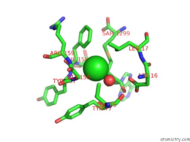
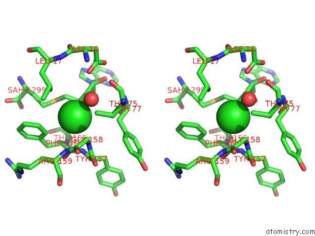
Chlorine binding site 1 out of 3 in 2v7t
Go back to
Chlorine binding site 1 out
of 3 in the X-Ray Crystal Structure of 5'-Fluorodeoxyadenosine Synthase S158G Mutant Complexed with S-Adenosyl-L-Homocysteine and Chloride Ion
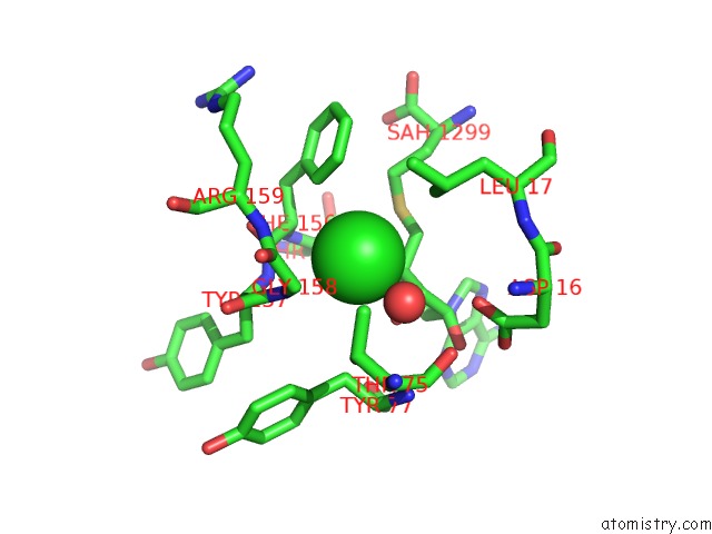
Mono view
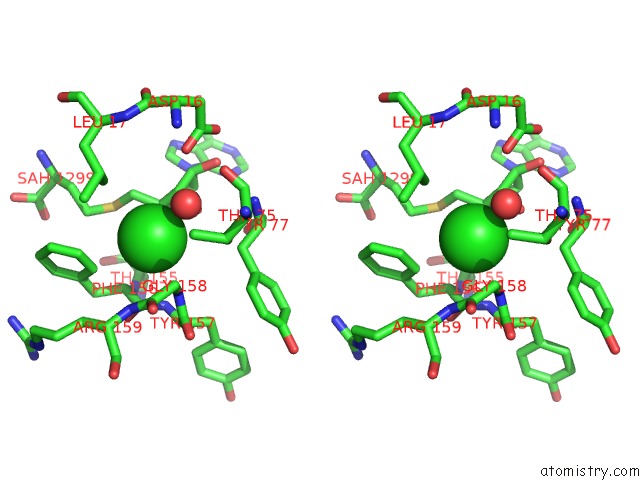
Stereo pair view
Mono view
Stereo pair view
A full contact list of Chlorine with other atoms in the Cl binding
site number 1 of X-Ray Crystal Structure of 5'-Fluorodeoxyadenosine Synthase S158G Mutant Complexed with S-Adenosyl-L-Homocysteine and Chloride Ion within 5.0Å range:
|
Chlorine binding site 2 out of 3 in 2v7t
Go back to
Chlorine binding site 2 out
of 3 in the X-Ray Crystal Structure of 5'-Fluorodeoxyadenosine Synthase S158G Mutant Complexed with S-Adenosyl-L-Homocysteine and Chloride Ion
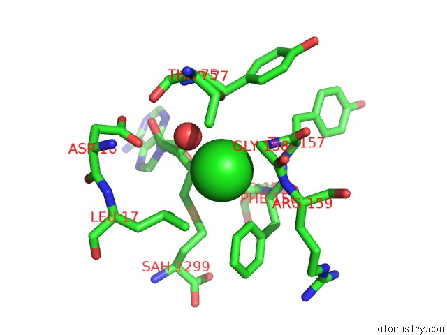
Mono view
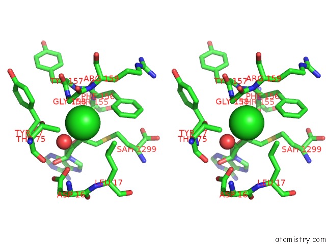
Stereo pair view
Mono view
Stereo pair view
A full contact list of Chlorine with other atoms in the Cl binding
site number 2 of X-Ray Crystal Structure of 5'-Fluorodeoxyadenosine Synthase S158G Mutant Complexed with S-Adenosyl-L-Homocysteine and Chloride Ion within 5.0Å range:
|
Chlorine binding site 3 out of 3 in 2v7t
Go back to
Chlorine binding site 3 out
of 3 in the X-Ray Crystal Structure of 5'-Fluorodeoxyadenosine Synthase S158G Mutant Complexed with S-Adenosyl-L-Homocysteine and Chloride Ion
Mono view
Stereo pair view
Mono view
Stereo pair view
A full contact list of Chlorine with other atoms in the Cl binding
site number 3 of X-Ray Crystal Structure of 5'-Fluorodeoxyadenosine Synthase S158G Mutant Complexed with S-Adenosyl-L-Homocysteine and Chloride Ion within 5.0Å range:
|
Reference:
X.Zhu,
D.A.Robinson,
A.R.Mcewan,
D.O'hagan,
J.H.Naismith.
Mechanism of Enzymatic Fluorination in Streptomyces Cattleya. J. Am. Chem. Soc. V. 129 14597 2007.
ISSN: ESSN 1520-5126
PubMed: 17985882
DOI: 10.1021/JA0731569
Page generated: Fri Jul 11 00:41:57 2025
ISSN: ESSN 1520-5126
PubMed: 17985882
DOI: 10.1021/JA0731569
Last articles
Fe in 2YXOFe in 2YRS
Fe in 2YXC
Fe in 2YNM
Fe in 2YVJ
Fe in 2YP1
Fe in 2YU2
Fe in 2YU1
Fe in 2YQB
Fe in 2YOO