Chlorine »
PDB 2v7f-2vfx »
2vd9 »
Chlorine in PDB 2vd9: The Crystal Structure of Alanine Racemase From Bacillus Anthracis (BA0252) with Bound L-Ala-P
Enzymatic activity of The Crystal Structure of Alanine Racemase From Bacillus Anthracis (BA0252) with Bound L-Ala-P
All present enzymatic activity of The Crystal Structure of Alanine Racemase From Bacillus Anthracis (BA0252) with Bound L-Ala-P:
5.1.1.1;
5.1.1.1;
Protein crystallography data
The structure of The Crystal Structure of Alanine Racemase From Bacillus Anthracis (BA0252) with Bound L-Ala-P, PDB code: 2vd9
was solved by
K.Au,
J.Ren,
T.S.Walter,
K.Harlos,
J.E.Nettleship,
R.J.Owens,
D.I.Stuart,
R.M.Esnouf,
Oxford Protein Production Facility (Oppf),
Structural Proteomics In Europe (Spine),
with X-Ray Crystallography technique. A brief refinement statistics is given in the table below:
| Resolution Low / High (Å) | 45.64 / 2.1 |
| Space group | P 21 21 21 |
| Cell size a, b, c (Å), α, β, γ (°) | 59.692, 96.504, 140.660, 90.00, 90.00, 90.00 |
| R / Rfree (%) | 18.8 / 23.9 |
Other elements in 2vd9:
The structure of The Crystal Structure of Alanine Racemase From Bacillus Anthracis (BA0252) with Bound L-Ala-P also contains other interesting chemical elements:
| Magnesium | (Mg) | 3 atoms |
Chlorine Binding Sites:
The binding sites of Chlorine atom in the The Crystal Structure of Alanine Racemase From Bacillus Anthracis (BA0252) with Bound L-Ala-P
(pdb code 2vd9). This binding sites where shown within
5.0 Angstroms radius around Chlorine atom.
In total 3 binding sites of Chlorine where determined in the The Crystal Structure of Alanine Racemase From Bacillus Anthracis (BA0252) with Bound L-Ala-P, PDB code: 2vd9:
Jump to Chlorine binding site number: 1; 2; 3;
In total 3 binding sites of Chlorine where determined in the The Crystal Structure of Alanine Racemase From Bacillus Anthracis (BA0252) with Bound L-Ala-P, PDB code: 2vd9:
Jump to Chlorine binding site number: 1; 2; 3;
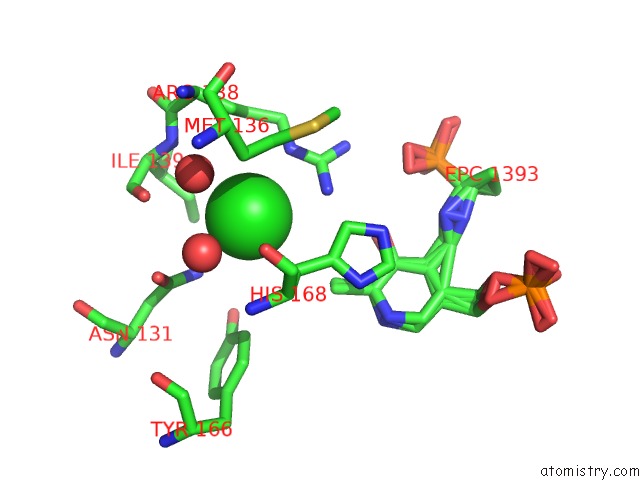
Chlorine binding site 1 out of 3 in 2vd9
Go back to
Chlorine binding site 1 out
of 3 in the The Crystal Structure of Alanine Racemase From Bacillus Anthracis (BA0252) with Bound L-Ala-P
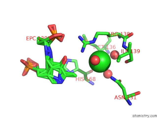
Mono view
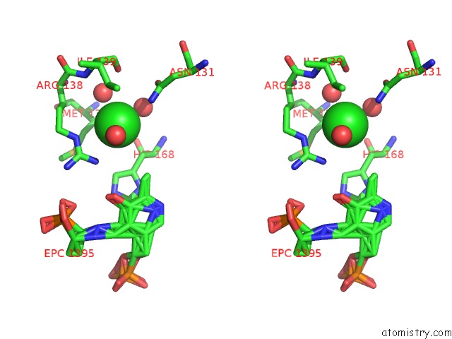
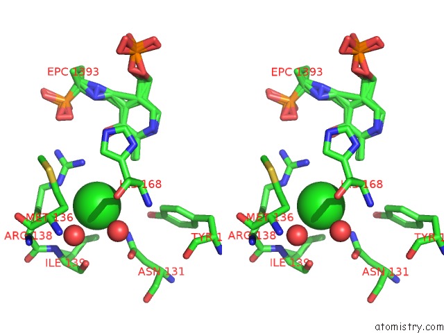
Stereo pair view
Mono view
Stereo pair view
A full contact list of Chlorine with other atoms in the Cl binding
site number 1 of The Crystal Structure of Alanine Racemase From Bacillus Anthracis (BA0252) with Bound L-Ala-P within 5.0Å range:
|
Chlorine binding site 2 out of 3 in 2vd9
Go back to
Chlorine binding site 2 out
of 3 in the The Crystal Structure of Alanine Racemase From Bacillus Anthracis (BA0252) with Bound L-Ala-P
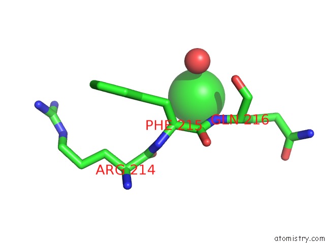
Mono view
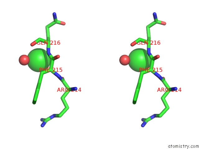
Stereo pair view
Mono view
Stereo pair view
A full contact list of Chlorine with other atoms in the Cl binding
site number 2 of The Crystal Structure of Alanine Racemase From Bacillus Anthracis (BA0252) with Bound L-Ala-P within 5.0Å range:
|
Chlorine binding site 3 out of 3 in 2vd9
Go back to
Chlorine binding site 3 out
of 3 in the The Crystal Structure of Alanine Racemase From Bacillus Anthracis (BA0252) with Bound L-Ala-P
Mono view
Stereo pair view
Mono view
Stereo pair view
A full contact list of Chlorine with other atoms in the Cl binding
site number 3 of The Crystal Structure of Alanine Racemase From Bacillus Anthracis (BA0252) with Bound L-Ala-P within 5.0Å range:
|
Reference:
K.Au,
J.Ren,
T.S.Walter,
K.Harlos,
J.E.Nettleship,
R.J.Owens,
D.I.Stuart,
R.M.Esnouf.
Structures of An Alanine Racemase From Bacillus Anthracis (BA0252) in the Presence and Absence of (R)-1-Aminoethylphosphonic Acid (L-Ala-P). Acta Crystallogr.,Sect.F V. 64 327 2008.
ISSN: ISSN 1744-3091
PubMed: 18453697
DOI: 10.1107/S1744309108007252
Page generated: Sat Jul 20 11:47:34 2024
ISSN: ISSN 1744-3091
PubMed: 18453697
DOI: 10.1107/S1744309108007252
Last articles
Zn in 9J0NZn in 9J0O
Zn in 9J0P
Zn in 9FJX
Zn in 9EKB
Zn in 9C0F
Zn in 9CAH
Zn in 9CH0
Zn in 9CH3
Zn in 9CH1