Chlorine »
PDB 2vo6-2vv8 »
2vrp »
Chlorine in PDB 2vrp: Structure of Rhodocytin
Protein crystallography data
The structure of Structure of Rhodocytin, PDB code: 2vrp
was solved by
A.A.Watson,
C.A.O'callaghan,
with X-Ray Crystallography technique. A brief refinement statistics is given in the table below:
| Resolution Low / High (Å) | 36.76 / 2.41 |
| Space group | I 2 2 2 |
| Cell size a, b, c (Å), α, β, γ (°) | 61.933, 89.368, 120.996, 90.00, 90.00, 90.00 |
| R / Rfree (%) | 20.1 / 27.2 |
Other elements in 2vrp:
The structure of Structure of Rhodocytin also contains other interesting chemical elements:
| Sodium | (Na) | 4 atoms |
Chlorine Binding Sites:
The binding sites of Chlorine atom in the Structure of Rhodocytin
(pdb code 2vrp). This binding sites where shown within
5.0 Angstroms radius around Chlorine atom.
In total 2 binding sites of Chlorine where determined in the Structure of Rhodocytin, PDB code: 2vrp:
Jump to Chlorine binding site number: 1; 2;
In total 2 binding sites of Chlorine where determined in the Structure of Rhodocytin, PDB code: 2vrp:
Jump to Chlorine binding site number: 1; 2;
Chlorine binding site 1 out of 2 in 2vrp
Go back to
Chlorine binding site 1 out
of 2 in the Structure of Rhodocytin
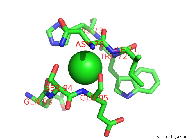
Mono view
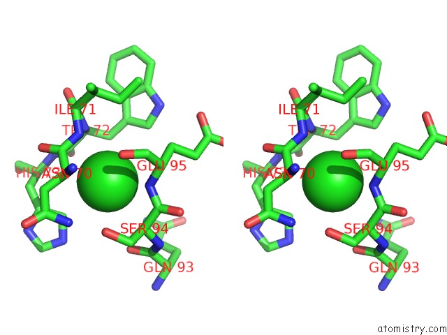
Stereo pair view
Mono view
Stereo pair view
A full contact list of Chlorine with other atoms in the Cl binding
site number 1 of Structure of Rhodocytin within 5.0Å range:
|
Chlorine binding site 2 out of 2 in 2vrp
Go back to
Chlorine binding site 2 out
of 2 in the Structure of Rhodocytin
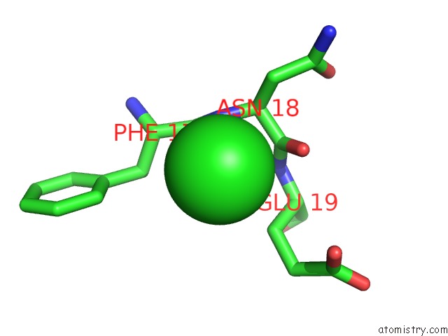
Mono view
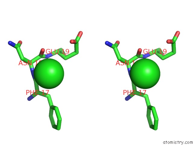
Stereo pair view
Mono view
Stereo pair view
A full contact list of Chlorine with other atoms in the Cl binding
site number 2 of Structure of Rhodocytin within 5.0Å range:
|
Reference:
A.A.Watson,
J.A.Eble,
C.A.O'callaghan.
Crystal Structure of Rhodocytin, A Ligand For the Platelet-Activating Receptor Clec-2. Protein Sci. V. 17 1611 2008.
ISSN: ISSN 0961-8368
PubMed: 18583525
DOI: 10.1110/PS.035568.108
Page generated: Sat Jul 20 12:12:09 2024
ISSN: ISSN 0961-8368
PubMed: 18583525
DOI: 10.1110/PS.035568.108
Last articles
Zn in 9MJ5Zn in 9HNW
Zn in 9G0L
Zn in 9FNE
Zn in 9DZN
Zn in 9E0I
Zn in 9D32
Zn in 9DAK
Zn in 8ZXC
Zn in 8ZUF