Chlorine »
PDB 2w2d-2wcp »
2wcg »
Chlorine in PDB 2wcg: X-Ray Structure of Acid-Beta-Glucosidase with N-Octyl(Cyclic Guanidine)-Nojirimycin in the Active Site
Enzymatic activity of X-Ray Structure of Acid-Beta-Glucosidase with N-Octyl(Cyclic Guanidine)-Nojirimycin in the Active Site
All present enzymatic activity of X-Ray Structure of Acid-Beta-Glucosidase with N-Octyl(Cyclic Guanidine)-Nojirimycin in the Active Site:
3.2.1.45;
3.2.1.45;
Protein crystallography data
The structure of X-Ray Structure of Acid-Beta-Glucosidase with N-Octyl(Cyclic Guanidine)-Nojirimycin in the Active Site, PDB code: 2wcg
was solved by
B.Brumshtein,
M.Aguilar,
M.I.Garcia-Moreno,
C.O.Mellet,
J.M.Garcia-Fernandez,
I.Silman,
Y.Shaaltiel,
D.Aviezer,
J.L.Sussman,
A.H.Futerman,
with X-Ray Crystallography technique. A brief refinement statistics is given in the table below:
| Resolution Low / High (Å) | 19.74 / 2.30 |
| Space group | P 1 21 1 |
| Cell size a, b, c (Å), α, β, γ (°) | 68.311, 96.827, 83.234, 90.00, 104.34, 90.00 |
| R / Rfree (%) | 13.5 / 19.4 |
Chlorine Binding Sites:
The binding sites of Chlorine atom in the X-Ray Structure of Acid-Beta-Glucosidase with N-Octyl(Cyclic Guanidine)-Nojirimycin in the Active Site
(pdb code 2wcg). This binding sites where shown within
5.0 Angstroms radius around Chlorine atom.
In total 2 binding sites of Chlorine where determined in the X-Ray Structure of Acid-Beta-Glucosidase with N-Octyl(Cyclic Guanidine)-Nojirimycin in the Active Site, PDB code: 2wcg:
Jump to Chlorine binding site number: 1; 2;
In total 2 binding sites of Chlorine where determined in the X-Ray Structure of Acid-Beta-Glucosidase with N-Octyl(Cyclic Guanidine)-Nojirimycin in the Active Site, PDB code: 2wcg:
Jump to Chlorine binding site number: 1; 2;
Chlorine binding site 1 out of 2 in 2wcg
Go back to
Chlorine binding site 1 out
of 2 in the X-Ray Structure of Acid-Beta-Glucosidase with N-Octyl(Cyclic Guanidine)-Nojirimycin in the Active Site
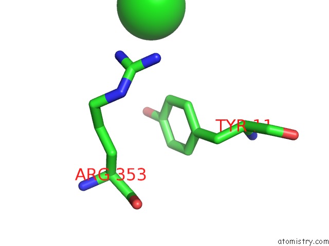
Mono view
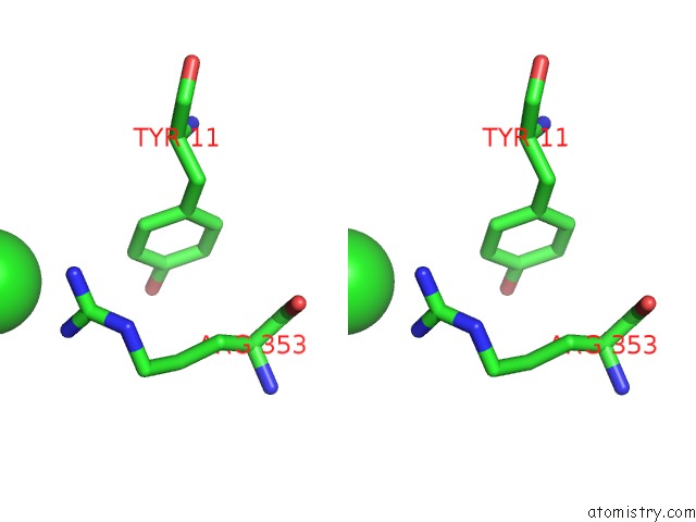
Stereo pair view
Mono view
Stereo pair view
A full contact list of Chlorine with other atoms in the Cl binding
site number 1 of X-Ray Structure of Acid-Beta-Glucosidase with N-Octyl(Cyclic Guanidine)-Nojirimycin in the Active Site within 5.0Å range:
|
Chlorine binding site 2 out of 2 in 2wcg
Go back to
Chlorine binding site 2 out
of 2 in the X-Ray Structure of Acid-Beta-Glucosidase with N-Octyl(Cyclic Guanidine)-Nojirimycin in the Active Site
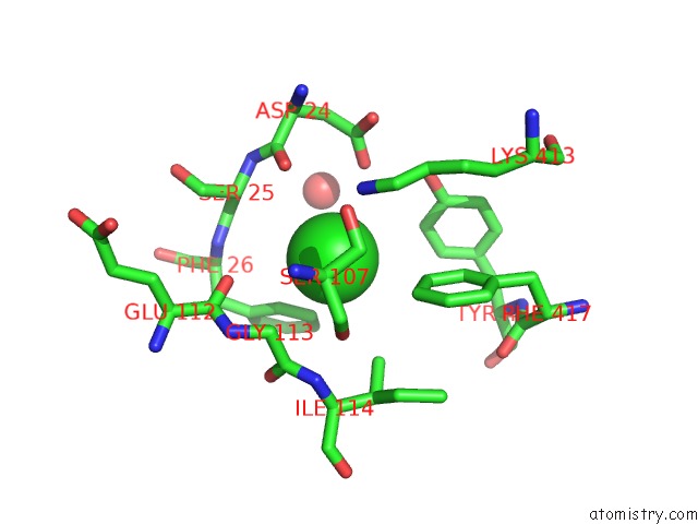
Mono view
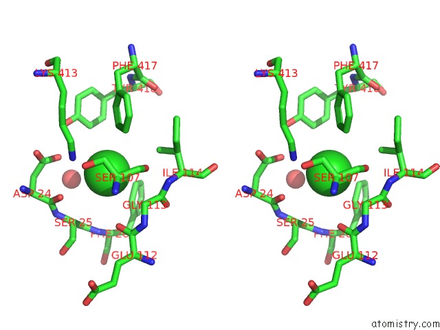
Stereo pair view
Mono view
Stereo pair view
A full contact list of Chlorine with other atoms in the Cl binding
site number 2 of X-Ray Structure of Acid-Beta-Glucosidase with N-Octyl(Cyclic Guanidine)-Nojirimycin in the Active Site within 5.0Å range:
|
Reference:
B.Brumshtein,
M.Aguilar-Moncayo,
M.I.Garcia-Moreno,
C.Ortiz Mellet,
J.M.Garcia Fernandez,
I.Silman,
Y.Shaaltiel,
D.Aviezer,
J.L.Sussman,
A.H.Futerman.
6-Amino-6-Deoxy-5,6-Di-N-(N'-Octyliminomethylidene) Nojirimycin: Synthesis, Biological Evaluation, and Crystal Structure in Complex with Acid Beta-Glucosidase. Chembiochem V. 10 1480 2009.
ISSN: ISSN 1439-4227
PubMed: 19437524
DOI: 10.1002/CBIC.200900142
Page generated: Sat Jul 20 12:40:36 2024
ISSN: ISSN 1439-4227
PubMed: 19437524
DOI: 10.1002/CBIC.200900142
Last articles
Zn in 9J0NZn in 9J0O
Zn in 9J0P
Zn in 9FJX
Zn in 9EKB
Zn in 9C0F
Zn in 9CAH
Zn in 9CH0
Zn in 9CH3
Zn in 9CH1