Chlorine »
PDB 2wcz-2wjp »
2wfj »
Chlorine in PDB 2wfj: Atomic Resolution Crystal Structure of the Ppiase Domain of Human Cyclophilin G in Complex with Cyclosporin A.
Enzymatic activity of Atomic Resolution Crystal Structure of the Ppiase Domain of Human Cyclophilin G in Complex with Cyclosporin A.
All present enzymatic activity of Atomic Resolution Crystal Structure of the Ppiase Domain of Human Cyclophilin G in Complex with Cyclosporin A.:
5.2.1.8;
5.2.1.8;
Protein crystallography data
The structure of Atomic Resolution Crystal Structure of the Ppiase Domain of Human Cyclophilin G in Complex with Cyclosporin A., PDB code: 2wfj
was solved by
C.M.Stegmann,
G.M.Sheldrick,
M.C.Wahl,
with X-Ray Crystallography technique. A brief refinement statistics is given in the table below:
| Resolution Low / High (Å) | 10.00 / 0.75 |
| Space group | P 21 21 21 |
| Cell size a, b, c (Å), α, β, γ (°) | 37.319, 64.913, 69.285, 90.00, 90.00, 90.00 |
| R / Rfree (%) | 11.1 / 12.9 |
Other elements in 2wfj:
The structure of Atomic Resolution Crystal Structure of the Ppiase Domain of Human Cyclophilin G in Complex with Cyclosporin A. also contains other interesting chemical elements:
| Magnesium | (Mg) | 2 atoms |
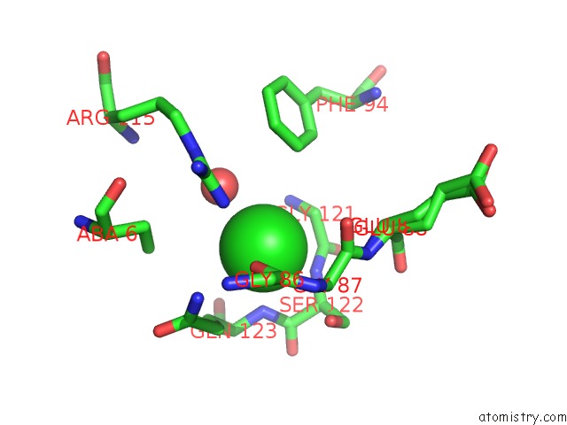
Chlorine Binding Sites:
The binding sites of Chlorine atom in the Atomic Resolution Crystal Structure of the Ppiase Domain of Human Cyclophilin G in Complex with Cyclosporin A.
(pdb code 2wfj). This binding sites where shown within
5.0 Angstroms radius around Chlorine atom.
In total only one binding site of Chlorine was determined in the Atomic Resolution Crystal Structure of the Ppiase Domain of Human Cyclophilin G in Complex with Cyclosporin A., PDB code: 2wfj:
In total only one binding site of Chlorine was determined in the Atomic Resolution Crystal Structure of the Ppiase Domain of Human Cyclophilin G in Complex with Cyclosporin A., PDB code: 2wfj:
Chlorine binding site 1 out of 1 in 2wfj
Go back to
Chlorine binding site 1 out
of 1 in the Atomic Resolution Crystal Structure of the Ppiase Domain of Human Cyclophilin G in Complex with Cyclosporin A.
Mono view
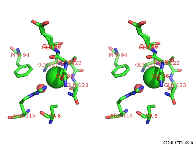
Stereo pair view
Mono view
Stereo pair view
A full contact list of Chlorine with other atoms in the Cl binding
site number 1 of Atomic Resolution Crystal Structure of the Ppiase Domain of Human Cyclophilin G in Complex with Cyclosporin A. within 5.0Å range:
|
Reference:
C.M.Stegmann,
D.Seeliger,
G.M.Sheldrick,
B.L.De Groot,
M.C.Wahl.
The Thermodynamic Influence of Trapped Water Molecules on A Protein-Ligand Interaction. Angew.Chem.Int.Ed.Engl. V. 48 5207 2009.
ISSN: ISSN 1433-7851
PubMed: 19499554
DOI: 10.1002/ANIE.200900481
Page generated: Sat Jul 20 12:47:14 2024
ISSN: ISSN 1433-7851
PubMed: 19499554
DOI: 10.1002/ANIE.200900481
Last articles
Zn in 9J0NZn in 9J0O
Zn in 9J0P
Zn in 9FJX
Zn in 9EKB
Zn in 9C0F
Zn in 9CAH
Zn in 9CH0
Zn in 9CH3
Zn in 9CH1