Chlorine »
PDB 2wcz-2wjp »
2wi8 »
Chlorine in PDB 2wi8: Crystal Structure of the Triscatecholate Siderophore Binding Protein Feua From Bacillus Subtilis
Protein crystallography data
The structure of Crystal Structure of the Triscatecholate Siderophore Binding Protein Feua From Bacillus Subtilis, PDB code: 2wi8
was solved by
F.Peuckert,
M.Miethke,
A.G.Albrecht,
L.-O.Essen,
M.A.Marahiel,
with X-Ray Crystallography technique. A brief refinement statistics is given in the table below:
| Resolution Low / High (Å) | 19.23 / 1.55 |
| Space group | P 41 21 2 |
| Cell size a, b, c (Å), α, β, γ (°) | 54.710, 54.710, 177.660, 90.00, 90.00, 90.00 |
| R / Rfree (%) | 18.178 / 21.544 |
Chlorine Binding Sites:
The binding sites of Chlorine atom in the Crystal Structure of the Triscatecholate Siderophore Binding Protein Feua From Bacillus Subtilis
(pdb code 2wi8). This binding sites where shown within
5.0 Angstroms radius around Chlorine atom.
In total 3 binding sites of Chlorine where determined in the Crystal Structure of the Triscatecholate Siderophore Binding Protein Feua From Bacillus Subtilis, PDB code: 2wi8:
Jump to Chlorine binding site number: 1; 2; 3;
In total 3 binding sites of Chlorine where determined in the Crystal Structure of the Triscatecholate Siderophore Binding Protein Feua From Bacillus Subtilis, PDB code: 2wi8:
Jump to Chlorine binding site number: 1; 2; 3;
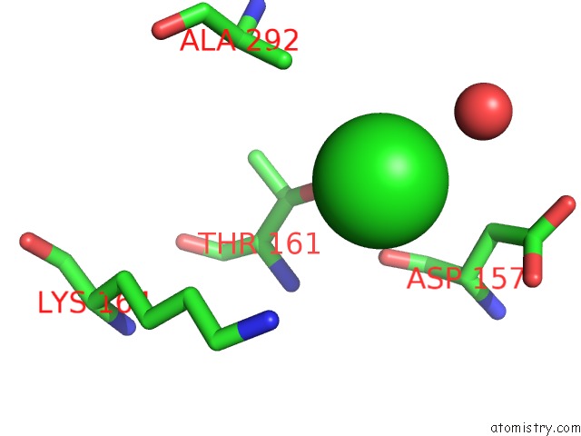
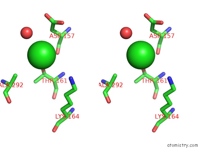
Chlorine binding site 1 out of 3 in 2wi8
Go back to
Chlorine binding site 1 out
of 3 in the Crystal Structure of the Triscatecholate Siderophore Binding Protein Feua From Bacillus Subtilis
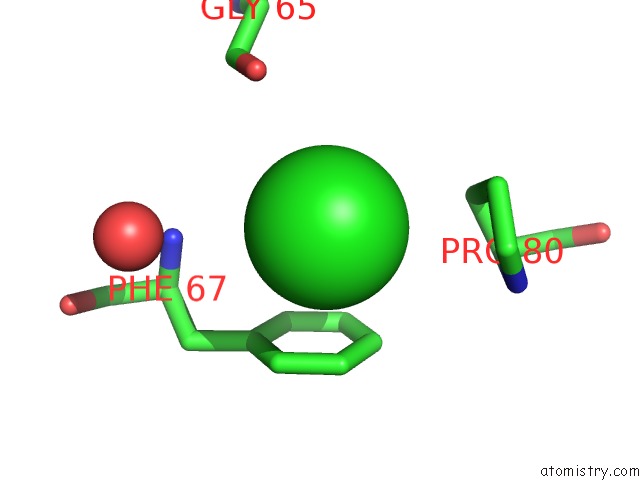
Mono view
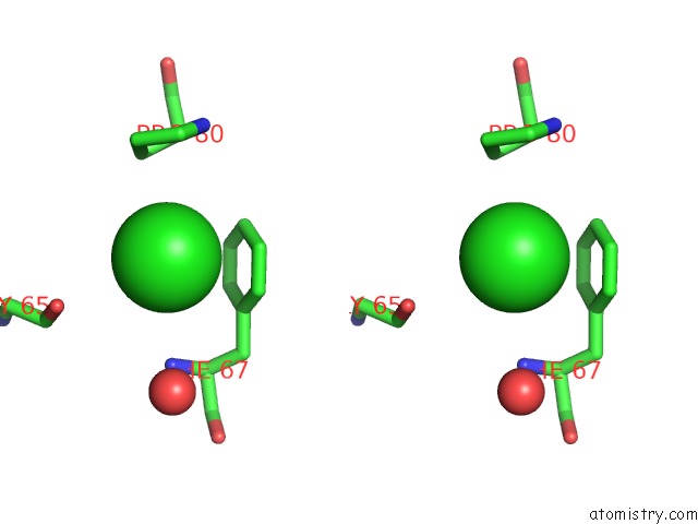
Stereo pair view
Mono view
Stereo pair view
A full contact list of Chlorine with other atoms in the Cl binding
site number 1 of Crystal Structure of the Triscatecholate Siderophore Binding Protein Feua From Bacillus Subtilis within 5.0Å range:
|
Chlorine binding site 2 out of 3 in 2wi8
Go back to
Chlorine binding site 2 out
of 3 in the Crystal Structure of the Triscatecholate Siderophore Binding Protein Feua From Bacillus Subtilis
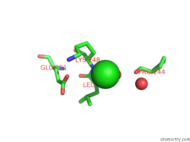
Mono view
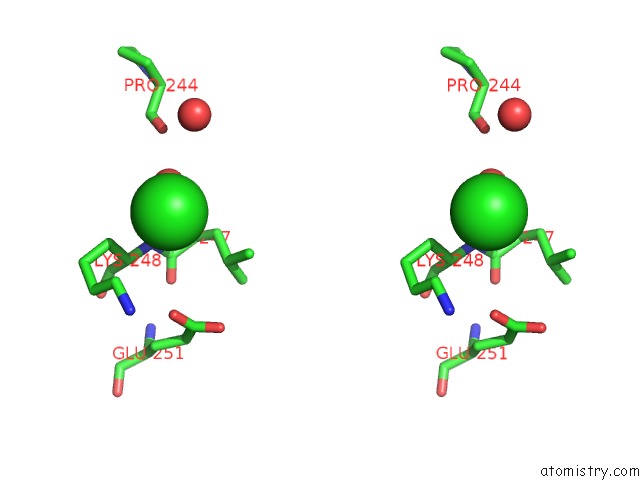
Stereo pair view
Mono view
Stereo pair view
A full contact list of Chlorine with other atoms in the Cl binding
site number 2 of Crystal Structure of the Triscatecholate Siderophore Binding Protein Feua From Bacillus Subtilis within 5.0Å range:
|
Chlorine binding site 3 out of 3 in 2wi8
Go back to
Chlorine binding site 3 out
of 3 in the Crystal Structure of the Triscatecholate Siderophore Binding Protein Feua From Bacillus Subtilis
Mono view
Stereo pair view
Mono view
Stereo pair view
A full contact list of Chlorine with other atoms in the Cl binding
site number 3 of Crystal Structure of the Triscatecholate Siderophore Binding Protein Feua From Bacillus Subtilis within 5.0Å range:
|
Reference:
F.Peuckert,
M.Miethke,
A.G.Albrecht,
L.-O.Essen,
M.A.Marahiel.
Structural Basis and Stereochemistry of Triscatecholate Siderophore Binding By Feua. Angew.Chem.Int.Ed.Engl. V. 48 7924 2009.
ISSN: ISSN 1433-7851
PubMed: 19746494
DOI: 10.1002/ANIE.200902495
Page generated: Sat Jul 20 12:54:23 2024
ISSN: ISSN 1433-7851
PubMed: 19746494
DOI: 10.1002/ANIE.200902495
Last articles
Zn in 9J0NZn in 9J0O
Zn in 9J0P
Zn in 9FJX
Zn in 9EKB
Zn in 9C0F
Zn in 9CAH
Zn in 9CH0
Zn in 9CH3
Zn in 9CH1