Chlorine »
PDB 2wps-2wzc »
2wsj »
Chlorine in PDB 2wsj: Crystal Structure of Single Point Mutant GLU71SER P-Coumaric Acid Decarboxylase
Protein crystallography data
The structure of Crystal Structure of Single Point Mutant GLU71SER P-Coumaric Acid Decarboxylase, PDB code: 2wsj
was solved by
H.Rodriguez,
I.Angulo,
B.De Las Rivas,
N.Campillo,
J.A.Paez,
R.Munoz,
J.M.Mancheno,
with X-Ray Crystallography technique. A brief refinement statistics is given in the table below:
| Resolution Low / High (Å) | 22.96 / 2.24 |
| Space group | C 1 2 1 |
| Cell size a, b, c (Å), α, β, γ (°) | 108.811, 52.756, 82.095, 90.00, 122.44, 90.00 |
| R / Rfree (%) | 17.8 / 25.9 |
Other elements in 2wsj:
The structure of Crystal Structure of Single Point Mutant GLU71SER P-Coumaric Acid Decarboxylase also contains other interesting chemical elements:
| Barium | (Ba) | 1 atom |
| Sodium | (Na) | 4 atoms |
Chlorine Binding Sites:
The binding sites of Chlorine atom in the Crystal Structure of Single Point Mutant GLU71SER P-Coumaric Acid Decarboxylase
(pdb code 2wsj). This binding sites where shown within
5.0 Angstroms radius around Chlorine atom.
In total only one binding site of Chlorine was determined in the Crystal Structure of Single Point Mutant GLU71SER P-Coumaric Acid Decarboxylase, PDB code: 2wsj:
In total only one binding site of Chlorine was determined in the Crystal Structure of Single Point Mutant GLU71SER P-Coumaric Acid Decarboxylase, PDB code: 2wsj:
Chlorine binding site 1 out of 1 in 2wsj
Go back to
Chlorine binding site 1 out
of 1 in the Crystal Structure of Single Point Mutant GLU71SER P-Coumaric Acid Decarboxylase
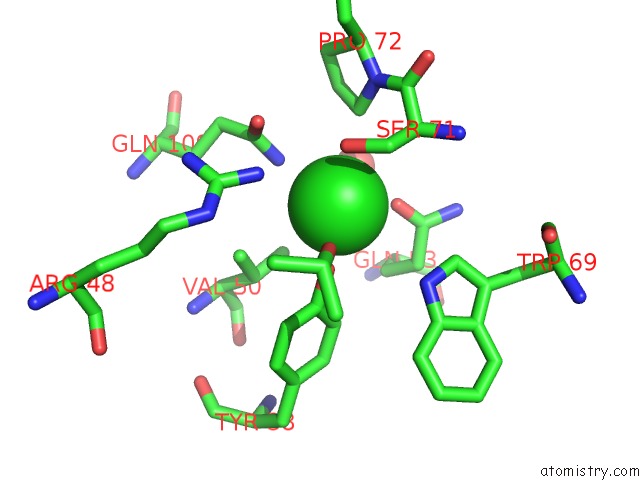
Mono view
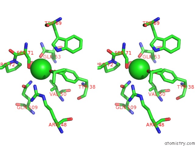
Stereo pair view
Mono view
Stereo pair view
A full contact list of Chlorine with other atoms in the Cl binding
site number 1 of Crystal Structure of Single Point Mutant GLU71SER P-Coumaric Acid Decarboxylase within 5.0Å range:
|
Reference:
H.Rodriguez,
I.Angulo,
B.De Las Rivas,
N.Campillo,
J.A.Paez,
R.Munoz,
J.M.Mancheno.
P-Coumaric Acid Decarboxylase From Lactobacillus Plantarum: Structural Insights Into the Active Site and Decarboxylation Catalytic Mechanism. Proteins V. 78 1662 2010.
ISSN: ISSN 0887-3585
PubMed: 20112419
DOI: 10.1002/PROT.22684
Page generated: Sat Jul 20 13:17:02 2024
ISSN: ISSN 0887-3585
PubMed: 20112419
DOI: 10.1002/PROT.22684
Last articles
Zn in 9J0NZn in 9J0O
Zn in 9J0P
Zn in 9FJX
Zn in 9EKB
Zn in 9C0F
Zn in 9CAH
Zn in 9CH0
Zn in 9CH3
Zn in 9CH1