Chlorine »
PDB 2wzd-2x8b »
2x2j »
Chlorine in PDB 2x2j: Crystal Structure of the Gracilariopsis Lemaneiformis Alpha- 1,4- Glucan Lyase with Deoxynojirimycin
Enzymatic activity of Crystal Structure of the Gracilariopsis Lemaneiformis Alpha- 1,4- Glucan Lyase with Deoxynojirimycin
All present enzymatic activity of Crystal Structure of the Gracilariopsis Lemaneiformis Alpha- 1,4- Glucan Lyase with Deoxynojirimycin:
4.2.2.13;
4.2.2.13;
Protein crystallography data
The structure of Crystal Structure of the Gracilariopsis Lemaneiformis Alpha- 1,4- Glucan Lyase with Deoxynojirimycin, PDB code: 2x2j
was solved by
H.J.Rozeboom,
S.Yu,
S.Madrid,
K.H.Kalk,
B.W.Dijkstra,
with X-Ray Crystallography technique. A brief refinement statistics is given in the table below:
| Resolution Low / High (Å) | 33.19 / 2.35 |
| Space group | P 1 21 1 |
| Cell size a, b, c (Å), α, β, γ (°) | 134.314, 91.707, 192.899, 90.00, 99.33, 90.00 |
| R / Rfree (%) | 23.4 / 29.1 |
Chlorine Binding Sites:
The binding sites of Chlorine atom in the Crystal Structure of the Gracilariopsis Lemaneiformis Alpha- 1,4- Glucan Lyase with Deoxynojirimycin
(pdb code 2x2j). This binding sites where shown within
5.0 Angstroms radius around Chlorine atom.
In total 4 binding sites of Chlorine where determined in the Crystal Structure of the Gracilariopsis Lemaneiformis Alpha- 1,4- Glucan Lyase with Deoxynojirimycin, PDB code: 2x2j:
Jump to Chlorine binding site number: 1; 2; 3; 4;
In total 4 binding sites of Chlorine where determined in the Crystal Structure of the Gracilariopsis Lemaneiformis Alpha- 1,4- Glucan Lyase with Deoxynojirimycin, PDB code: 2x2j:
Jump to Chlorine binding site number: 1; 2; 3; 4;
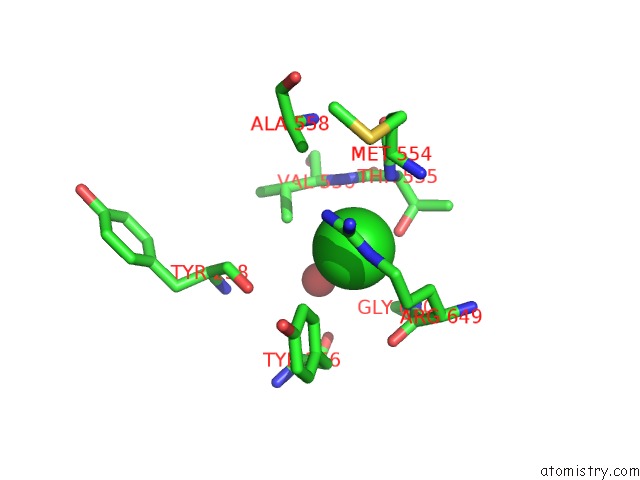
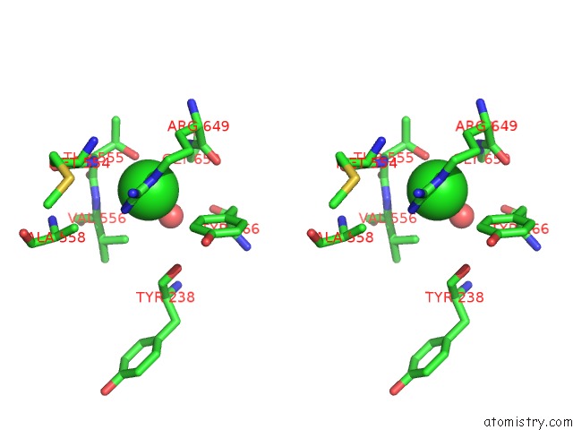
Chlorine binding site 1 out of 4 in 2x2j
Go back to
Chlorine binding site 1 out
of 4 in the Crystal Structure of the Gracilariopsis Lemaneiformis Alpha- 1,4- Glucan Lyase with Deoxynojirimycin
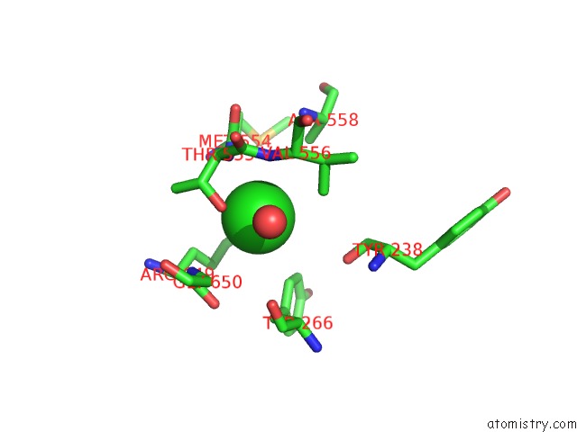
Mono view
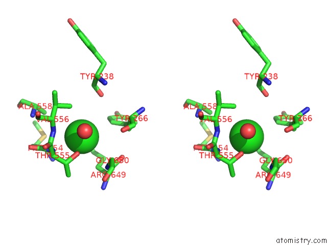
Stereo pair view
Mono view
Stereo pair view
A full contact list of Chlorine with other atoms in the Cl binding
site number 1 of Crystal Structure of the Gracilariopsis Lemaneiformis Alpha- 1,4- Glucan Lyase with Deoxynojirimycin within 5.0Å range:
|
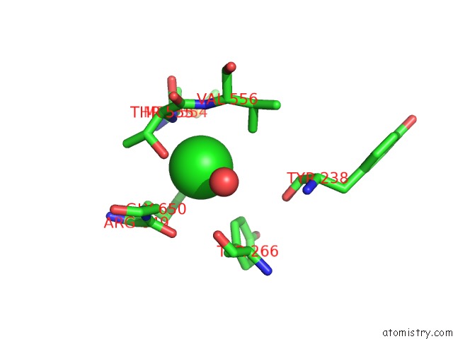
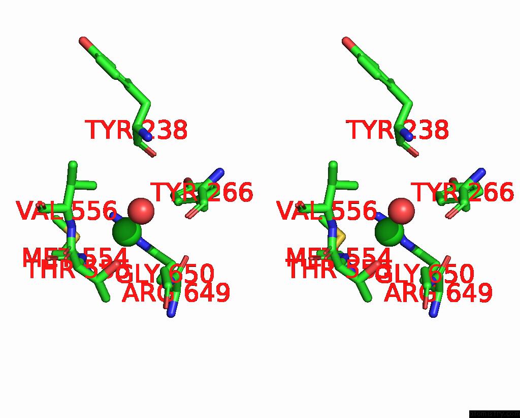
Chlorine binding site 2 out of 4 in 2x2j
Go back to
Chlorine binding site 2 out
of 4 in the Crystal Structure of the Gracilariopsis Lemaneiformis Alpha- 1,4- Glucan Lyase with Deoxynojirimycin
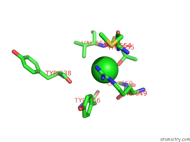
Mono view
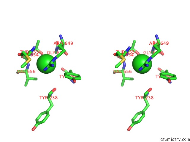
Stereo pair view
Mono view
Stereo pair view
A full contact list of Chlorine with other atoms in the Cl binding
site number 2 of Crystal Structure of the Gracilariopsis Lemaneiformis Alpha- 1,4- Glucan Lyase with Deoxynojirimycin within 5.0Å range:
|
Chlorine binding site 3 out of 4 in 2x2j
Go back to
Chlorine binding site 3 out
of 4 in the Crystal Structure of the Gracilariopsis Lemaneiformis Alpha- 1,4- Glucan Lyase with Deoxynojirimycin
Mono view
Stereo pair view
Mono view
Stereo pair view
A full contact list of Chlorine with other atoms in the Cl binding
site number 3 of Crystal Structure of the Gracilariopsis Lemaneiformis Alpha- 1,4- Glucan Lyase with Deoxynojirimycin within 5.0Å range:
|
Chlorine binding site 4 out of 4 in 2x2j
Go back to
Chlorine binding site 4 out
of 4 in the Crystal Structure of the Gracilariopsis Lemaneiformis Alpha- 1,4- Glucan Lyase with Deoxynojirimycin
Mono view
Stereo pair view
Mono view
Stereo pair view
A full contact list of Chlorine with other atoms in the Cl binding
site number 4 of Crystal Structure of the Gracilariopsis Lemaneiformis Alpha- 1,4- Glucan Lyase with Deoxynojirimycin within 5.0Å range:
|
Reference:
H.J.Rozeboom,
S.Yu,
S.Madrid,
K.H.Kalk,
R.Zhang,
B.W.Dijkstra.
Crystal Structure of Alpha-1,4-Glucan Lyase, A Unique Glycoside Hydrolase Family Member with A Novel Catalytic Mechanism. J.Biol.Chem. V. 288 26764 2013.
ISSN: ISSN 0021-9258
PubMed: 23902768
DOI: 10.1074/JBC.M113.485896
Page generated: Sat Jul 20 13:29:07 2024
ISSN: ISSN 0021-9258
PubMed: 23902768
DOI: 10.1074/JBC.M113.485896
Last articles
Zn in 9J0NZn in 9J0O
Zn in 9J0P
Zn in 9FJX
Zn in 9EKB
Zn in 9C0F
Zn in 9CAH
Zn in 9CH0
Zn in 9CH3
Zn in 9CH1