Chlorine »
PDB 2wzd-2x8b »
2x5g »
Chlorine in PDB 2x5g: Crystal Structure of the ORF131L51M Mutant From Sulfolobus Islandicus Rudivirus 1
Protein crystallography data
The structure of Crystal Structure of the ORF131L51M Mutant From Sulfolobus Islandicus Rudivirus 1, PDB code: 2x5g
was solved by
M.Oke,
L.G.Carter,
K.A.Johnson,
H.Liu,
S.A.Mcmahon,
J.H.Naismith,
M.F.White,
with X-Ray Crystallography technique. A brief refinement statistics is given in the table below:
| Resolution Low / High (Å) | 25.31 / 2.00 |
| Space group | P 31 2 1 |
| Cell size a, b, c (Å), α, β, γ (°) | 58.453, 58.453, 68.113, 90.00, 90.00, 120.00 |
| R / Rfree (%) | 21 / 24.9 |
Chlorine Binding Sites:
The binding sites of Chlorine atom in the Crystal Structure of the ORF131L51M Mutant From Sulfolobus Islandicus Rudivirus 1
(pdb code 2x5g). This binding sites where shown within
5.0 Angstroms radius around Chlorine atom.
In total 3 binding sites of Chlorine where determined in the Crystal Structure of the ORF131L51M Mutant From Sulfolobus Islandicus Rudivirus 1, PDB code: 2x5g:
Jump to Chlorine binding site number: 1; 2; 3;
In total 3 binding sites of Chlorine where determined in the Crystal Structure of the ORF131L51M Mutant From Sulfolobus Islandicus Rudivirus 1, PDB code: 2x5g:
Jump to Chlorine binding site number: 1; 2; 3;
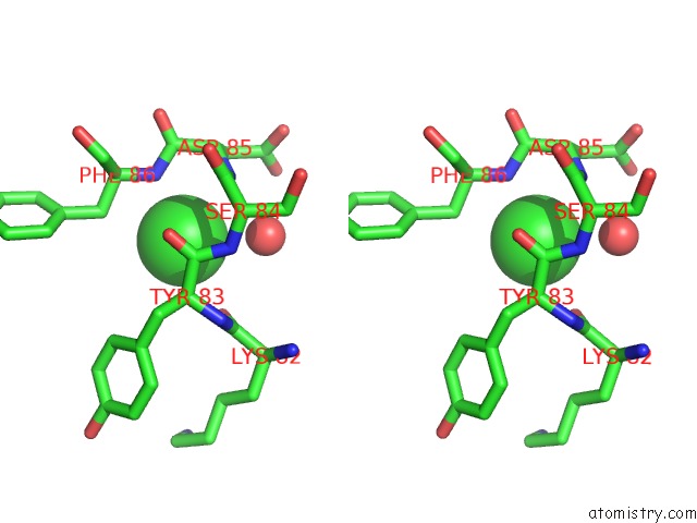
Chlorine binding site 1 out of 3 in 2x5g
Go back to
Chlorine binding site 1 out
of 3 in the Crystal Structure of the ORF131L51M Mutant From Sulfolobus Islandicus Rudivirus 1

Mono view

Stereo pair view
Mono view
Stereo pair view
A full contact list of Chlorine with other atoms in the Cl binding
site number 1 of Crystal Structure of the ORF131L51M Mutant From Sulfolobus Islandicus Rudivirus 1 within 5.0Å range:
|
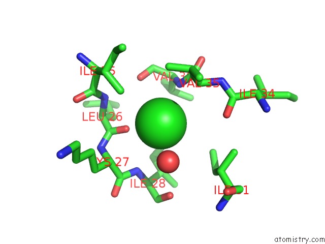
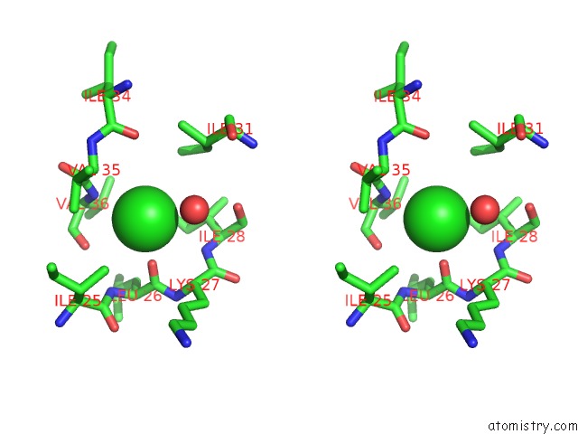
Chlorine binding site 2 out of 3 in 2x5g
Go back to
Chlorine binding site 2 out
of 3 in the Crystal Structure of the ORF131L51M Mutant From Sulfolobus Islandicus Rudivirus 1

Mono view
Stereo pair view
Mono view
Stereo pair view
A full contact list of Chlorine with other atoms in the Cl binding
site number 2 of Crystal Structure of the ORF131L51M Mutant From Sulfolobus Islandicus Rudivirus 1 within 5.0Å range:
|
Chlorine binding site 3 out of 3 in 2x5g
Go back to
Chlorine binding site 3 out
of 3 in the Crystal Structure of the ORF131L51M Mutant From Sulfolobus Islandicus Rudivirus 1
Mono view
Stereo pair view
Mono view
Stereo pair view
A full contact list of Chlorine with other atoms in the Cl binding
site number 3 of Crystal Structure of the ORF131L51M Mutant From Sulfolobus Islandicus Rudivirus 1 within 5.0Å range:
|
Reference:
M.Oke,
L.G.Carter,
K.A.Johnson,
H.Liu,
S.A.Mcmahon,
X.Yan,
M.Kerou,
N.D.Weikart,
N.Kadi,
M.A.Sheikh,
S.Schmelz,
M.Dorward,
M.Zawadzki,
C.Cozens,
H.Falconer,
H.Powers,
I.M.Overton,
C.A.J.Van Niekerk,
X.Peng,
P.Patel,
R.A.Garrett,
D.Prangishvili,
C.H.Botting,
P.J.Coote,
D.T.F.Dryden,
G.J.Barton,
U.Schwarz-Linek,
G.L.Challis,
G.L.Taylor,
M.F.White,
J.H.Naismith.
The Scottish Structural Proteomics Facility: Targets, Methods and Outputs. J.Struct.Funct.Genom. V. 11 167 2010.
ISSN: ISSN 1345-711X
PubMed: 20419351
DOI: 10.1007/S10969-010-9090-Y
Page generated: Sat Jul 20 13:30:07 2024
ISSN: ISSN 1345-711X
PubMed: 20419351
DOI: 10.1007/S10969-010-9090-Y
Last articles
Zn in 9J0NZn in 9J0O
Zn in 9J0P
Zn in 9FJX
Zn in 9EKB
Zn in 9C0F
Zn in 9CAH
Zn in 9CH0
Zn in 9CH3
Zn in 9CH1