Chlorine »
PDB 2xmd-2xs6 »
2xr6 »
Chlorine in PDB 2xr6: Crystal Structure of the Complex of the Carbohydrate Recognition Domain of Human Dc-Sign with Pseudo Trimannoside Mimic.
Protein crystallography data
The structure of Crystal Structure of the Complex of the Carbohydrate Recognition Domain of Human Dc-Sign with Pseudo Trimannoside Mimic., PDB code: 2xr6
was solved by
M.Thepaut,
I.Suitkeviciute,
S.Sattin,
J.Reina,
A.Bernardi,
F.Fieschi,
with X-Ray Crystallography technique. A brief refinement statistics is given in the table below:
| Resolution Low / High (Å) | 50.44 / 1.35 |
| Space group | P 43 21 2 |
| Cell size a, b, c (Å), α, β, γ (°) | 71.330, 71.330, 52.666, 90.00, 90.00, 90.00 |
| R / Rfree (%) | 14.7 / 16.8 |
Other elements in 2xr6:
The structure of Crystal Structure of the Complex of the Carbohydrate Recognition Domain of Human Dc-Sign with Pseudo Trimannoside Mimic. also contains other interesting chemical elements:
| Calcium | (Ca) | 3 atoms |
Chlorine Binding Sites:
The binding sites of Chlorine atom in the Crystal Structure of the Complex of the Carbohydrate Recognition Domain of Human Dc-Sign with Pseudo Trimannoside Mimic.
(pdb code 2xr6). This binding sites where shown within
5.0 Angstroms radius around Chlorine atom.
In total 3 binding sites of Chlorine where determined in the Crystal Structure of the Complex of the Carbohydrate Recognition Domain of Human Dc-Sign with Pseudo Trimannoside Mimic., PDB code: 2xr6:
Jump to Chlorine binding site number: 1; 2; 3;
In total 3 binding sites of Chlorine where determined in the Crystal Structure of the Complex of the Carbohydrate Recognition Domain of Human Dc-Sign with Pseudo Trimannoside Mimic., PDB code: 2xr6:
Jump to Chlorine binding site number: 1; 2; 3;
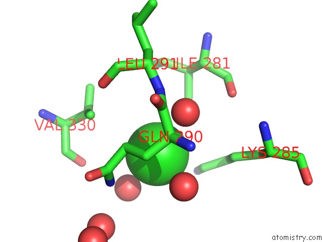
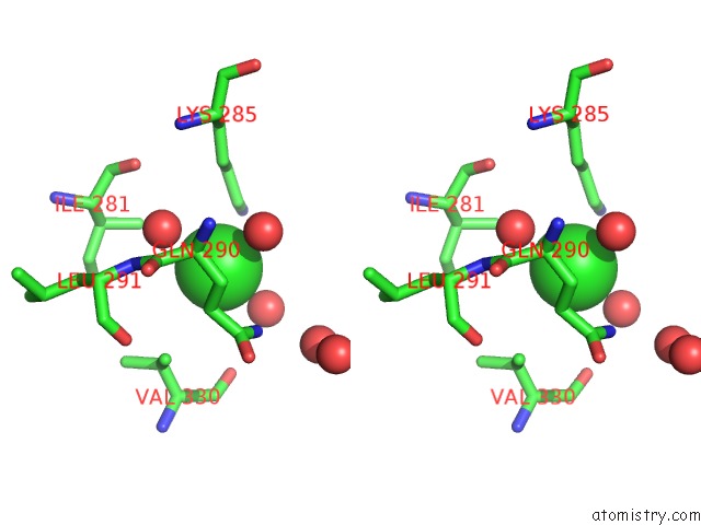
Chlorine binding site 1 out of 3 in 2xr6
Go back to
Chlorine binding site 1 out
of 3 in the Crystal Structure of the Complex of the Carbohydrate Recognition Domain of Human Dc-Sign with Pseudo Trimannoside Mimic.
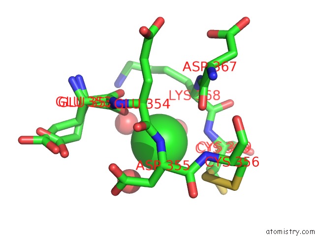
Mono view
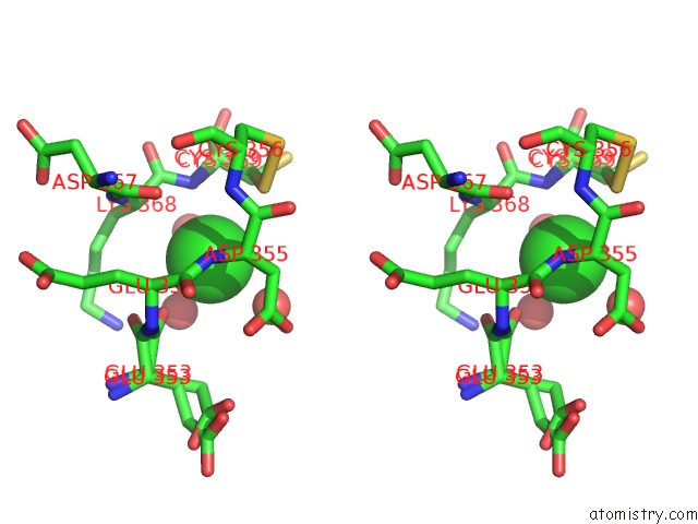
Stereo pair view
Mono view
Stereo pair view
A full contact list of Chlorine with other atoms in the Cl binding
site number 1 of Crystal Structure of the Complex of the Carbohydrate Recognition Domain of Human Dc-Sign with Pseudo Trimannoside Mimic. within 5.0Å range:
|
Chlorine binding site 2 out of 3 in 2xr6
Go back to
Chlorine binding site 2 out
of 3 in the Crystal Structure of the Complex of the Carbohydrate Recognition Domain of Human Dc-Sign with Pseudo Trimannoside Mimic.
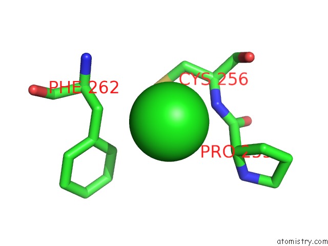
Mono view
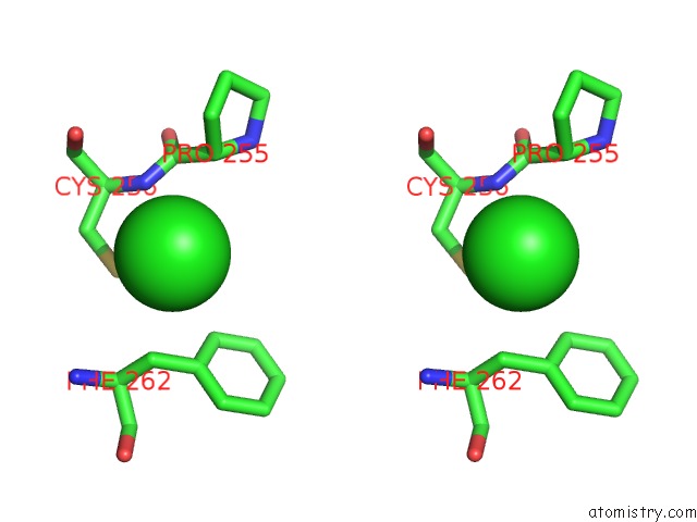
Stereo pair view
Mono view
Stereo pair view
A full contact list of Chlorine with other atoms in the Cl binding
site number 2 of Crystal Structure of the Complex of the Carbohydrate Recognition Domain of Human Dc-Sign with Pseudo Trimannoside Mimic. within 5.0Å range:
|
Chlorine binding site 3 out of 3 in 2xr6
Go back to
Chlorine binding site 3 out
of 3 in the Crystal Structure of the Complex of the Carbohydrate Recognition Domain of Human Dc-Sign with Pseudo Trimannoside Mimic.
Mono view
Stereo pair view
Mono view
Stereo pair view
A full contact list of Chlorine with other atoms in the Cl binding
site number 3 of Crystal Structure of the Complex of the Carbohydrate Recognition Domain of Human Dc-Sign with Pseudo Trimannoside Mimic. within 5.0Å range:
|
Reference:
I.Sutkeviciute,
M.Thepaut,
S.Sattin,
A.Berzi,
J.Mcgeagh,
S.Grudinin,
J.Weiser,
A.Le Roy,
J.J.Reina,
J.Rojo,
M.Clerici,
A.Bernardi,
C.Ebel,
F.Fieschi.
Unique Dc-Sign Clustering Activity of A Small Glycomimetic: A Lesson For Ligand Design. Acs Chem.Biol. V. 9 1377 2014.
ISSN: ISSN 1554-8929
PubMed: 24749535
DOI: 10.1021/CB500054H
Page generated: Sat Jul 20 14:08:46 2024
ISSN: ISSN 1554-8929
PubMed: 24749535
DOI: 10.1021/CB500054H
Last articles
Zn in 9J0NZn in 9J0O
Zn in 9J0P
Zn in 9FJX
Zn in 9EKB
Zn in 9C0F
Zn in 9CAH
Zn in 9CH0
Zn in 9CH3
Zn in 9CH1