Chlorine »
PDB 2xsa-2xzk »
2xtz »
Chlorine in PDB 2xtz: Crystal Structure of the G Alpha Protein ATGPA1 From Arabidopsis Thaliana
Enzymatic activity of Crystal Structure of the G Alpha Protein ATGPA1 From Arabidopsis Thaliana
All present enzymatic activity of Crystal Structure of the G Alpha Protein ATGPA1 From Arabidopsis Thaliana:
3.6.5.1;
3.6.5.1;
Protein crystallography data
The structure of Crystal Structure of the G Alpha Protein ATGPA1 From Arabidopsis Thaliana, PDB code: 2xtz
was solved by
J.C.Jones,
J.W.Duffy,
M.Machius,
B.R.S.Temple,
H.G.Dohlman,
A.M.Jones,
with X-Ray Crystallography technique. A brief refinement statistics is given in the table below:
| Resolution Low / High (Å) | 96.03 / 2.34 |
| Space group | P 21 21 21 |
| Cell size a, b, c (Å), α, β, γ (°) | 67.130, 119.347, 161.725, 90.00, 90.00, 90.00 |
| R / Rfree (%) | 21.1 / 25.4 |
Other elements in 2xtz:
The structure of Crystal Structure of the G Alpha Protein ATGPA1 From Arabidopsis Thaliana also contains other interesting chemical elements:
| Magnesium | (Mg) | 3 atoms |
Chlorine Binding Sites:
The binding sites of Chlorine atom in the Crystal Structure of the G Alpha Protein ATGPA1 From Arabidopsis Thaliana
(pdb code 2xtz). This binding sites where shown within
5.0 Angstroms radius around Chlorine atom.
In total 4 binding sites of Chlorine where determined in the Crystal Structure of the G Alpha Protein ATGPA1 From Arabidopsis Thaliana, PDB code: 2xtz:
Jump to Chlorine binding site number: 1; 2; 3; 4;
In total 4 binding sites of Chlorine where determined in the Crystal Structure of the G Alpha Protein ATGPA1 From Arabidopsis Thaliana, PDB code: 2xtz:
Jump to Chlorine binding site number: 1; 2; 3; 4;
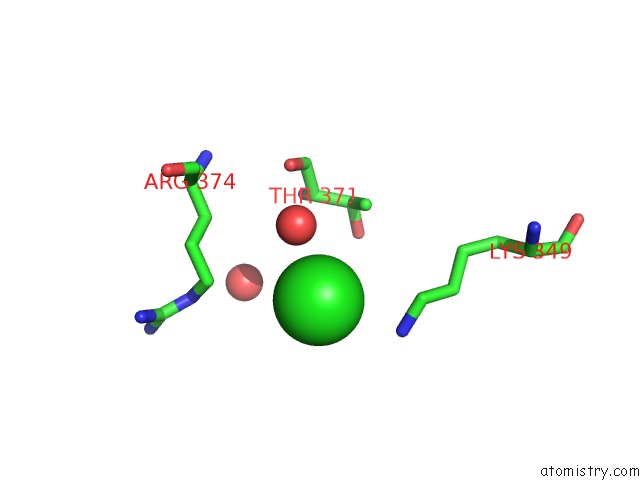
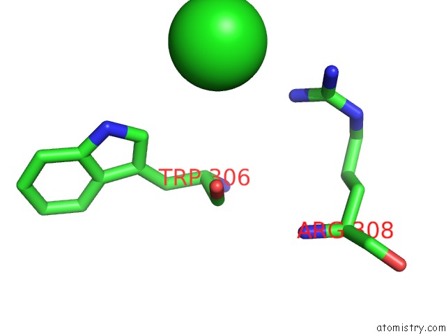
Chlorine binding site 1 out of 4 in 2xtz
Go back to
Chlorine binding site 1 out
of 4 in the Crystal Structure of the G Alpha Protein ATGPA1 From Arabidopsis Thaliana
Mono view
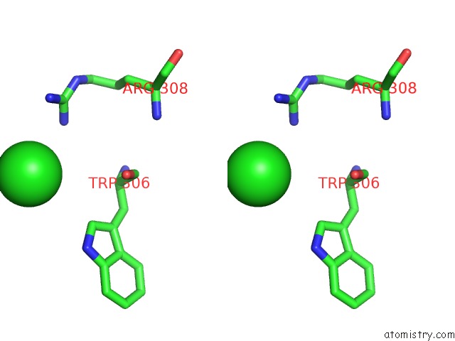
Stereo pair view
Mono view
Stereo pair view
A full contact list of Chlorine with other atoms in the Cl binding
site number 1 of Crystal Structure of the G Alpha Protein ATGPA1 From Arabidopsis Thaliana within 5.0Å range:
|
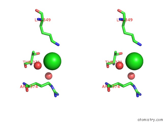
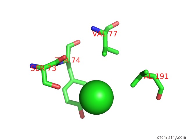
Chlorine binding site 2 out of 4 in 2xtz
Go back to
Chlorine binding site 2 out
of 4 in the Crystal Structure of the G Alpha Protein ATGPA1 From Arabidopsis Thaliana
Mono view
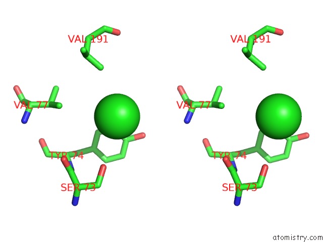
Stereo pair view
Mono view
Stereo pair view
A full contact list of Chlorine with other atoms in the Cl binding
site number 2 of Crystal Structure of the G Alpha Protein ATGPA1 From Arabidopsis Thaliana within 5.0Å range:
|
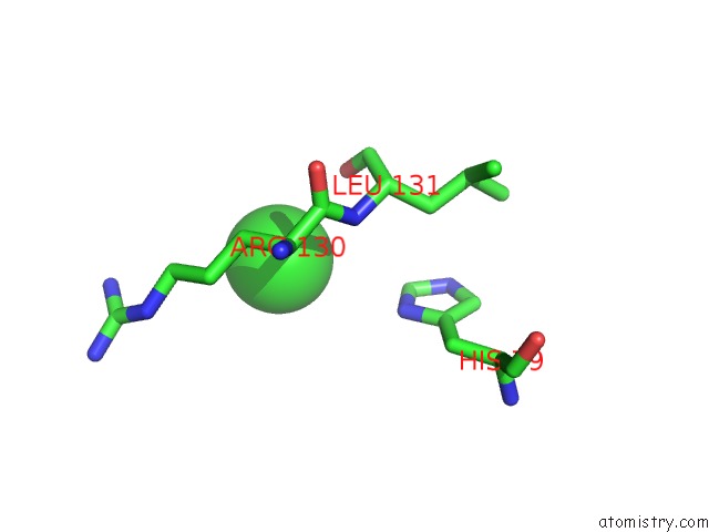
Chlorine binding site 3 out of 4 in 2xtz
Go back to
Chlorine binding site 3 out
of 4 in the Crystal Structure of the G Alpha Protein ATGPA1 From Arabidopsis Thaliana
Mono view
Stereo pair view
Mono view
Stereo pair view
A full contact list of Chlorine with other atoms in the Cl binding
site number 3 of Crystal Structure of the G Alpha Protein ATGPA1 From Arabidopsis Thaliana within 5.0Å range:
|
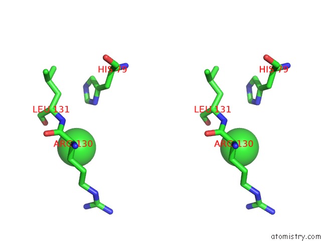
Chlorine binding site 4 out of 4 in 2xtz
Go back to
Chlorine binding site 4 out
of 4 in the Crystal Structure of the G Alpha Protein ATGPA1 From Arabidopsis Thaliana
Mono view
Stereo pair view
Mono view
Stereo pair view
A full contact list of Chlorine with other atoms in the Cl binding
site number 4 of Crystal Structure of the G Alpha Protein ATGPA1 From Arabidopsis Thaliana within 5.0Å range:
|
Reference:
J.C.Jones,
J.W.Duffy,
M.Machius,
B.R.S.Temple,
H.G.Dohlman,
A.M.Jones.
The Crystal Structure of A Self-Activating G Protein Alpha Subunit Reveals Its Distinct Mechanism of Signal Initiation Sci.Signal. V. 159 RA8 2011.
ISSN: ESSN 1937-9145
PubMed: 21304159
DOI: 10.1126/SCISIGNAL.2001446
Page generated: Sat Jul 20 14:10:47 2024
ISSN: ESSN 1937-9145
PubMed: 21304159
DOI: 10.1126/SCISIGNAL.2001446
Last articles
Zn in 9J0NZn in 9J0O
Zn in 9J0P
Zn in 9FJX
Zn in 9EKB
Zn in 9C0F
Zn in 9CAH
Zn in 9CH0
Zn in 9CH3
Zn in 9CH1