Chlorine »
PDB 2xzr-2y58 »
2y1d »
Chlorine in PDB 2y1d: X-Ray Structure of 1-Deoxy-D-Xylulose 5-Phosphate Reductoisomerase, Dxr, RV2870C, From Mycobacterium Tuberculosis, in Complex with A 3,4- Dichlorophenyl-Substituted Fosmidomycin Analogue and Manganese.
Enzymatic activity of X-Ray Structure of 1-Deoxy-D-Xylulose 5-Phosphate Reductoisomerase, Dxr, RV2870C, From Mycobacterium Tuberculosis, in Complex with A 3,4- Dichlorophenyl-Substituted Fosmidomycin Analogue and Manganese.
All present enzymatic activity of X-Ray Structure of 1-Deoxy-D-Xylulose 5-Phosphate Reductoisomerase, Dxr, RV2870C, From Mycobacterium Tuberculosis, in Complex with A 3,4- Dichlorophenyl-Substituted Fosmidomycin Analogue and Manganese.:
1.1.1.267;
1.1.1.267;
Protein crystallography data
The structure of X-Ray Structure of 1-Deoxy-D-Xylulose 5-Phosphate Reductoisomerase, Dxr, RV2870C, From Mycobacterium Tuberculosis, in Complex with A 3,4- Dichlorophenyl-Substituted Fosmidomycin Analogue and Manganese., PDB code: 2y1d
was solved by
L.M.Henriksson,
A.M.S.Larsson,
T.Bergfors,
C.Bjorkelid,
T.Unge,
S.L.Mowbray,
T.A.Jones,
with X-Ray Crystallography technique. A brief refinement statistics is given in the table below:
| Resolution Low / High (Å) | 40.00 / 2.05 |
| Space group | P 1 21 1 |
| Cell size a, b, c (Å), α, β, γ (°) | 69.622, 67.318, 84.301, 90.00, 107.38, 90.00 |
| R / Rfree (%) | 20.9 / 26.1 |
Other elements in 2y1d:
The structure of X-Ray Structure of 1-Deoxy-D-Xylulose 5-Phosphate Reductoisomerase, Dxr, RV2870C, From Mycobacterium Tuberculosis, in Complex with A 3,4- Dichlorophenyl-Substituted Fosmidomycin Analogue and Manganese. also contains other interesting chemical elements:
| Manganese | (Mn) | 2 atoms |
Chlorine Binding Sites:
The binding sites of Chlorine atom in the X-Ray Structure of 1-Deoxy-D-Xylulose 5-Phosphate Reductoisomerase, Dxr, RV2870C, From Mycobacterium Tuberculosis, in Complex with A 3,4- Dichlorophenyl-Substituted Fosmidomycin Analogue and Manganese.
(pdb code 2y1d). This binding sites where shown within
5.0 Angstroms radius around Chlorine atom.
In total 2 binding sites of Chlorine where determined in the X-Ray Structure of 1-Deoxy-D-Xylulose 5-Phosphate Reductoisomerase, Dxr, RV2870C, From Mycobacterium Tuberculosis, in Complex with A 3,4- Dichlorophenyl-Substituted Fosmidomycin Analogue and Manganese., PDB code: 2y1d:
Jump to Chlorine binding site number: 1; 2;
Chlorine binding site 1 out of 2 in 2y1d
Go back to
Chlorine binding site 1 out
of 2 in the X-Ray Structure of 1-Deoxy-D-Xylulose 5-Phosphate Reductoisomerase, Dxr, RV2870C, From Mycobacterium Tuberculosis, in Complex with A 3,4- Dichlorophenyl-Substituted Fosmidomycin Analogue and Manganese.
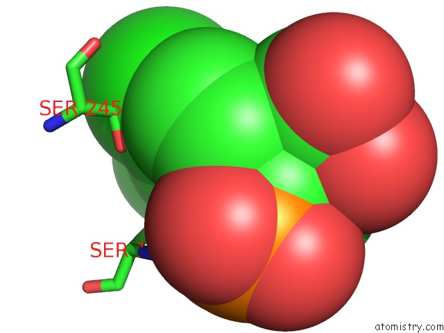
Mono view
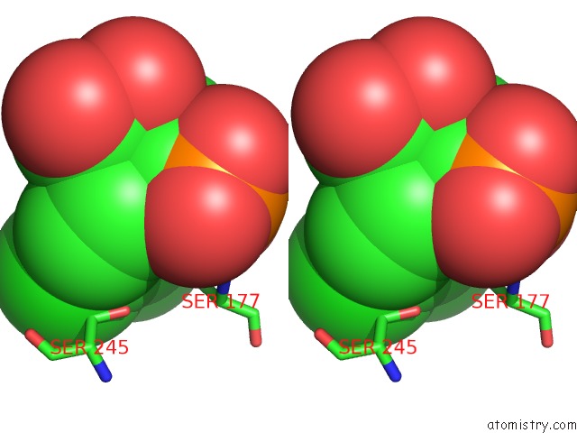
Stereo pair view
Mono view
Stereo pair view
A full contact list of Chlorine with other atoms in the Cl binding
site number 1 of X-Ray Structure of 1-Deoxy-D-Xylulose 5-Phosphate Reductoisomerase, Dxr, RV2870C, From Mycobacterium Tuberculosis, in Complex with A 3,4- Dichlorophenyl-Substituted Fosmidomycin Analogue and Manganese. within 5.0Å range:
|
Chlorine binding site 2 out of 2 in 2y1d
Go back to
Chlorine binding site 2 out
of 2 in the X-Ray Structure of 1-Deoxy-D-Xylulose 5-Phosphate Reductoisomerase, Dxr, RV2870C, From Mycobacterium Tuberculosis, in Complex with A 3,4- Dichlorophenyl-Substituted Fosmidomycin Analogue and Manganese.
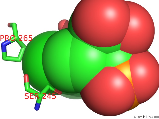
Mono view
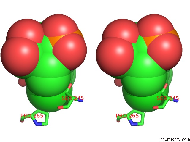
Stereo pair view
Mono view
Stereo pair view
A full contact list of Chlorine with other atoms in the Cl binding
site number 2 of X-Ray Structure of 1-Deoxy-D-Xylulose 5-Phosphate Reductoisomerase, Dxr, RV2870C, From Mycobacterium Tuberculosis, in Complex with A 3,4- Dichlorophenyl-Substituted Fosmidomycin Analogue and Manganese. within 5.0Å range:
|
Reference:
M.Andaloussi,
L.M.Henriksson,
A.Wieckowska,
M.Lindh,
C.Bjorkelid,
A.M.S.Larsson,
H.Iyer,
B.R.Srinivasa,
T.Bergfors,
T.Unge,
S.L.Mowbray,
M.Larhed,
T.A.Jones,
A.Karlen.
Design, Synthesis and X-Ray Crystallographic Studies of Alpha-Aryl Substituted Fosmidomycin Analogues As Inhibitors of Mycobacterium Tuberculosis 1-Deoxy-D-Xylulose-5-Phosphate Reductoisomerase J.Med.Chem V. 54 4964 2011.
ISSN: ISSN 0022-2623
PubMed: 21678907
DOI: 10.1021/JM2000085
Page generated: Sat Jul 20 14:22:04 2024
ISSN: ISSN 0022-2623
PubMed: 21678907
DOI: 10.1021/JM2000085
Last articles
Zn in 9J0NZn in 9J0O
Zn in 9J0P
Zn in 9FJX
Zn in 9EKB
Zn in 9C0F
Zn in 9CAH
Zn in 9CH0
Zn in 9CH3
Zn in 9CH1