Chlorine »
PDB 2y5f-2yc5 »
2y6o »
Chlorine in PDB 2y6o: Crystal Structure of EPHA4 Kinase Domain in Complex with Dasatinib.
Enzymatic activity of Crystal Structure of EPHA4 Kinase Domain in Complex with Dasatinib.
All present enzymatic activity of Crystal Structure of EPHA4 Kinase Domain in Complex with Dasatinib.:
2.7.10.1;
2.7.10.1;
Protein crystallography data
The structure of Crystal Structure of EPHA4 Kinase Domain in Complex with Dasatinib., PDB code: 2y6o
was solved by
C.J.A.Farenc,
P.H.N.Celie,
G.Siegal,
with X-Ray Crystallography technique. A brief refinement statistics is given in the table below:
| Resolution Low / High (Å) | 19.588 / 1.54 |
| Space group | P 21 21 21 |
| Cell size a, b, c (Å), α, β, γ (°) | 32.412, 91.636, 98.346, 90.00, 90.00, 90.00 |
| R / Rfree (%) | 18.01 / 20.05 |
Chlorine Binding Sites:
The binding sites of Chlorine atom in the Crystal Structure of EPHA4 Kinase Domain in Complex with Dasatinib.
(pdb code 2y6o). This binding sites where shown within
5.0 Angstroms radius around Chlorine atom.
In total only one binding site of Chlorine was determined in the Crystal Structure of EPHA4 Kinase Domain in Complex with Dasatinib., PDB code: 2y6o:
In total only one binding site of Chlorine was determined in the Crystal Structure of EPHA4 Kinase Domain in Complex with Dasatinib., PDB code: 2y6o:
Chlorine binding site 1 out of 1 in 2y6o
Go back to
Chlorine binding site 1 out
of 1 in the Crystal Structure of EPHA4 Kinase Domain in Complex with Dasatinib.
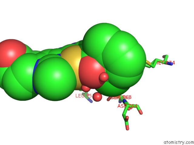
Mono view
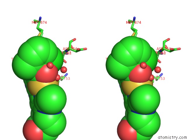
Stereo pair view
Mono view
Stereo pair view
A full contact list of Chlorine with other atoms in the Cl binding
site number 1 of Crystal Structure of EPHA4 Kinase Domain in Complex with Dasatinib. within 5.0Å range:
|
Reference:
C.J.A.Farenc,
L.Hameetman,
W.Zoutman,
C.P.Tensen,
G.Siegal.
Crystal Structure of the EPHA4 Protein Tyrosine Kinase Domain in the Apo- and Dasatinib-Bound State. Febs Lett. V. 585 3593 2011.
ISSN: ISSN 0014-5793
PubMed: 22036717
DOI: 10.1016/J.FEBSLET.2011.10.028
Page generated: Fri Jul 11 02:31:32 2025
ISSN: ISSN 0014-5793
PubMed: 22036717
DOI: 10.1016/J.FEBSLET.2011.10.028
Last articles
Cl in 3Q0PCl in 3Q0D
Cl in 3Q03
Cl in 3Q02
Cl in 3PXP
Cl in 3PZH
Cl in 3PYK
Cl in 3PYH
Cl in 3PYC
Cl in 3PWM