Chlorine »
PDB 2yjb-2yzr »
2yve »
Chlorine in PDB 2yve: Crystal Structure of the Methylene Blue-Bound Form of the Multi-Drug Binding Transcriptional Repressor Cgmr
Protein crystallography data
The structure of Crystal Structure of the Methylene Blue-Bound Form of the Multi-Drug Binding Transcriptional Repressor Cgmr, PDB code: 2yve
was solved by
H.Itou,
Y.Shirakihara,
I.Tanaka,
with X-Ray Crystallography technique. A brief refinement statistics is given in the table below:
| Resolution Low / High (Å) | 9.97 / 1.40 |
| Space group | P 21 21 21 |
| Cell size a, b, c (Å), α, β, γ (°) | 60.110, 67.360, 101.850, 90.00, 90.00, 90.00 |
| R / Rfree (%) | 20.3 / 22.3 |
Chlorine Binding Sites:
The binding sites of Chlorine atom in the Crystal Structure of the Methylene Blue-Bound Form of the Multi-Drug Binding Transcriptional Repressor Cgmr
(pdb code 2yve). This binding sites where shown within
5.0 Angstroms radius around Chlorine atom.
In total only one binding site of Chlorine was determined in the Crystal Structure of the Methylene Blue-Bound Form of the Multi-Drug Binding Transcriptional Repressor Cgmr, PDB code: 2yve:
In total only one binding site of Chlorine was determined in the Crystal Structure of the Methylene Blue-Bound Form of the Multi-Drug Binding Transcriptional Repressor Cgmr, PDB code: 2yve:
Chlorine binding site 1 out of 1 in 2yve
Go back to
Chlorine binding site 1 out
of 1 in the Crystal Structure of the Methylene Blue-Bound Form of the Multi-Drug Binding Transcriptional Repressor Cgmr
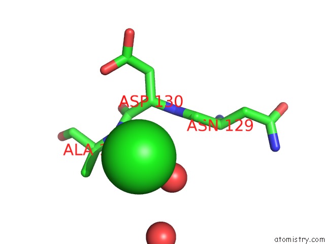
Mono view
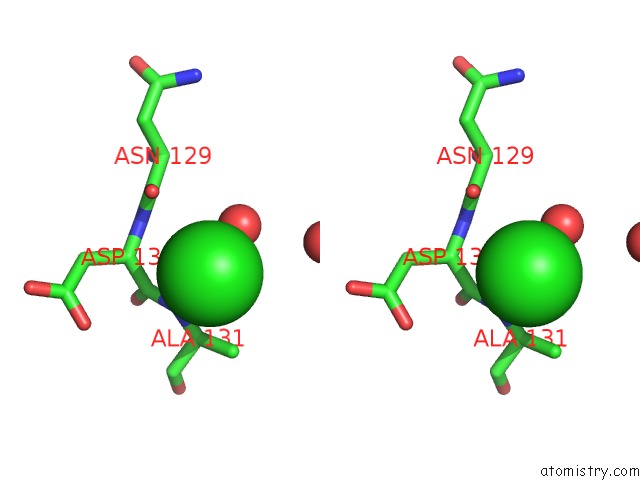
Stereo pair view
Mono view
Stereo pair view
A full contact list of Chlorine with other atoms in the Cl binding
site number 1 of Crystal Structure of the Methylene Blue-Bound Form of the Multi-Drug Binding Transcriptional Repressor Cgmr within 5.0Å range:
|
Reference:
H.Itou,
N.Watanabe,
M.Yao,
Y.Shirakihara,
I.Tanaka.
Crystal Structures of the Multidrug Binding Repressor Corynebacteriumglutamicum Cgmr in Complex with Inducers and with An Operator J.Mol.Biol. V. 403 174 2010.
ISSN: ISSN 0022-2836
PubMed: 20691702
DOI: 10.1016/J.JMB.2010.07.042
Page generated: Sat Jul 20 15:17:26 2024
ISSN: ISSN 0022-2836
PubMed: 20691702
DOI: 10.1016/J.JMB.2010.07.042
Last articles
Zn in 9J0NZn in 9J0O
Zn in 9J0P
Zn in 9FJX
Zn in 9EKB
Zn in 9C0F
Zn in 9CAH
Zn in 9CH0
Zn in 9CH3
Zn in 9CH1