Chlorine »
PDB 2z0g-2zgd »
2z2k »
Chlorine in PDB 2z2k: Crystal Structure of Peptidyl-Trna Hydrolase From Mycobacterium Tuberculosis
Enzymatic activity of Crystal Structure of Peptidyl-Trna Hydrolase From Mycobacterium Tuberculosis
All present enzymatic activity of Crystal Structure of Peptidyl-Trna Hydrolase From Mycobacterium Tuberculosis:
3.1.1.29;
3.1.1.29;
Protein crystallography data
The structure of Crystal Structure of Peptidyl-Trna Hydrolase From Mycobacterium Tuberculosis, PDB code: 2z2k
was solved by
M.Selvaraj,
S.Roy,
N.S.Singh,
R.Sangeetha,
U.Varshney,
M.Vijayan,
with X-Ray Crystallography technique. A brief refinement statistics is given in the table below:
| Resolution Low / High (Å) | 19.01 / 2.50 |
| Space group | P 21 21 21 |
| Cell size a, b, c (Å), α, β, γ (°) | 35.840, 57.060, 72.590, 90.00, 90.00, 90.00 |
| R / Rfree (%) | 19.8 / 24.1 |
Chlorine Binding Sites:
The binding sites of Chlorine atom in the Crystal Structure of Peptidyl-Trna Hydrolase From Mycobacterium Tuberculosis
(pdb code 2z2k). This binding sites where shown within
5.0 Angstroms radius around Chlorine atom.
In total only one binding site of Chlorine was determined in the Crystal Structure of Peptidyl-Trna Hydrolase From Mycobacterium Tuberculosis, PDB code: 2z2k:
In total only one binding site of Chlorine was determined in the Crystal Structure of Peptidyl-Trna Hydrolase From Mycobacterium Tuberculosis, PDB code: 2z2k:
Chlorine binding site 1 out of 1 in 2z2k
Go back to
Chlorine binding site 1 out
of 1 in the Crystal Structure of Peptidyl-Trna Hydrolase From Mycobacterium Tuberculosis
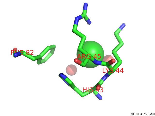
Mono view
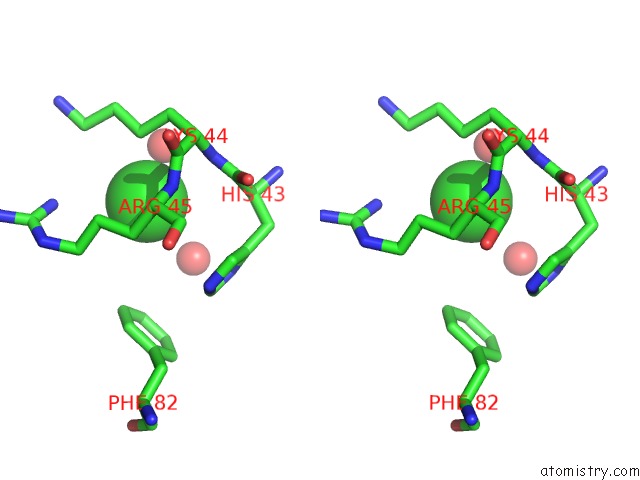
Stereo pair view
Mono view
Stereo pair view
A full contact list of Chlorine with other atoms in the Cl binding
site number 1 of Crystal Structure of Peptidyl-Trna Hydrolase From Mycobacterium Tuberculosis within 5.0Å range:
|
Reference:
M.Selvaraj,
S.Roy,
N.S.Singh,
R.Sangeetha,
U.Varshney,
M.Vijayan.
Structural Plasticity and Enzyme Action: Crystal Structures of Mycobacterium Tuberculosis Peptidyl-Trna Hydrolase J.Mol.Biol. V. 372 186 2007.
ISSN: ISSN 0022-2836
PubMed: 17619020
DOI: 10.1016/J.JMB.2007.06.053
Page generated: Fri Jul 11 02:49:50 2025
ISSN: ISSN 0022-2836
PubMed: 17619020
DOI: 10.1016/J.JMB.2007.06.053
Last articles
Cl in 3MVXCl in 3MVY
Cl in 3MVU
Cl in 3MUX
Cl in 3MVT
Cl in 3MV6
Cl in 3MTN
Cl in 3MUJ
Cl in 3MTU
Cl in 3MTH