Chlorine »
PDB 2zhp-3a34 »
2zm4 »
Chlorine in PDB 2zm4: Crystal Structure of Imidazo Quinoxaline 1 Bound to the Kinase Domain of Human Lck, Activated Form (Auto- Phosphorylated on TYR394)
Enzymatic activity of Crystal Structure of Imidazo Quinoxaline 1 Bound to the Kinase Domain of Human Lck, Activated Form (Auto- Phosphorylated on TYR394)
All present enzymatic activity of Crystal Structure of Imidazo Quinoxaline 1 Bound to the Kinase Domain of Human Lck, Activated Form (Auto- Phosphorylated on TYR394):
2.7.10.2;
2.7.10.2;
Protein crystallography data
The structure of Crystal Structure of Imidazo Quinoxaline 1 Bound to the Kinase Domain of Human Lck, Activated Form (Auto- Phosphorylated on TYR394), PDB code: 2zm4
was solved by
E.Tsuji,
with X-Ray Crystallography technique. A brief refinement statistics is given in the table below:
| Resolution Low / High (Å) | 15.00 / 2.70 |
| Space group | P 21 21 21 |
| Cell size a, b, c (Å), α, β, γ (°) | 42.591, 73.807, 92.159, 90.00, 90.00, 90.00 |
| R / Rfree (%) | 17.6 / 27.4 |
Chlorine Binding Sites:
The binding sites of Chlorine atom in the Crystal Structure of Imidazo Quinoxaline 1 Bound to the Kinase Domain of Human Lck, Activated Form (Auto- Phosphorylated on TYR394)
(pdb code 2zm4). This binding sites where shown within
5.0 Angstroms radius around Chlorine atom.
In total only one binding site of Chlorine was determined in the Crystal Structure of Imidazo Quinoxaline 1 Bound to the Kinase Domain of Human Lck, Activated Form (Auto- Phosphorylated on TYR394), PDB code: 2zm4:
In total only one binding site of Chlorine was determined in the Crystal Structure of Imidazo Quinoxaline 1 Bound to the Kinase Domain of Human Lck, Activated Form (Auto- Phosphorylated on TYR394), PDB code: 2zm4:
Chlorine binding site 1 out of 1 in 2zm4
Go back to
Chlorine binding site 1 out
of 1 in the Crystal Structure of Imidazo Quinoxaline 1 Bound to the Kinase Domain of Human Lck, Activated Form (Auto- Phosphorylated on TYR394)
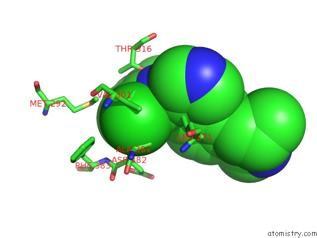
Mono view
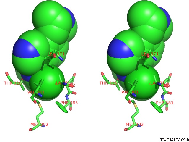
Stereo pair view
Mono view
Stereo pair view
A full contact list of Chlorine with other atoms in the Cl binding
site number 1 of Crystal Structure of Imidazo Quinoxaline 1 Bound to the Kinase Domain of Human Lck, Activated Form (Auto- Phosphorylated on TYR394) within 5.0Å range:
|
Reference:
T.Ozawa,
E.Tsuji,
M.Ozawa,
C.Handa,
H.Mukaiyama,
T.Nishimura,
S.Kobayashi,
K.Okazaki.
The Importance of Ch/Pi Hydrogen Bonds in Rational Drug Design: An Ab Initio Fragment Molecular Orbital Study to Leukocyte-Specific Protein Tyrosine (Lck) Kinase Bioorg.Med.Chem. V. 16 10311 2008.
ISSN: ISSN 0968-0896
PubMed: 18977146
DOI: 10.1016/J.BMC.2008.10.041
Page generated: Sat Jul 20 15:34:33 2024
ISSN: ISSN 0968-0896
PubMed: 18977146
DOI: 10.1016/J.BMC.2008.10.041
Last articles
Zn in 9J0NZn in 9J0O
Zn in 9J0P
Zn in 9FJX
Zn in 9EKB
Zn in 9C0F
Zn in 9CAH
Zn in 9CH0
Zn in 9CH3
Zn in 9CH1