Chlorine »
PDB 3a4p-3akf »
3ab9 »
Chlorine in PDB 3ab9: Crystal Structure of Lipoylated E. Coli H-Protein (Reduced Form)
Protein crystallography data
The structure of Crystal Structure of Lipoylated E. Coli H-Protein (Reduced Form), PDB code: 3ab9
was solved by
K.Okamura-Ikeda,
N.Maita,
with X-Ray Crystallography technique. A brief refinement statistics is given in the table below:
| Resolution Low / High (Å) | 20.00 / 1.65 |
| Space group | P 43 21 2 |
| Cell size a, b, c (Å), α, β, γ (°) | 60.255, 60.255, 68.586, 90.00, 90.00, 90.00 |
| R / Rfree (%) | 20.4 / 21.6 |
Other elements in 3ab9:
The structure of Crystal Structure of Lipoylated E. Coli H-Protein (Reduced Form) also contains other interesting chemical elements:
| Calcium | (Ca) | 1 atom |
Chlorine Binding Sites:
The binding sites of Chlorine atom in the Crystal Structure of Lipoylated E. Coli H-Protein (Reduced Form)
(pdb code 3ab9). This binding sites where shown within
5.0 Angstroms radius around Chlorine atom.
In total only one binding site of Chlorine was determined in the Crystal Structure of Lipoylated E. Coli H-Protein (Reduced Form), PDB code: 3ab9:
In total only one binding site of Chlorine was determined in the Crystal Structure of Lipoylated E. Coli H-Protein (Reduced Form), PDB code: 3ab9:
Chlorine binding site 1 out of 1 in 3ab9
Go back to
Chlorine binding site 1 out
of 1 in the Crystal Structure of Lipoylated E. Coli H-Protein (Reduced Form)
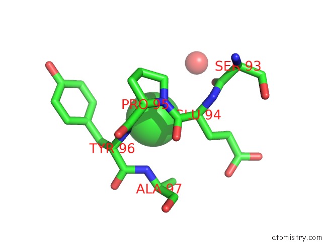
Mono view
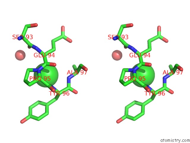
Stereo pair view
Mono view
Stereo pair view
A full contact list of Chlorine with other atoms in the Cl binding
site number 1 of Crystal Structure of Lipoylated E. Coli H-Protein (Reduced Form) within 5.0Å range:
|
Reference:
K.Okamura-Ikeda,
H.Hosaka,
N.Maita,
K.Fujiwara,
A.C.Yoshizawa,
A.Nakagawa,
H.Taniguchi.
Crystal Structure of Aminomethyltransferase in Complex with Dihydrolipoyl-H-Protein of the Glycine Cleavage System: Implications For Recognition of Lipoyl Protein Substrate, Disease-Related Mutations, and Reaction Mechanism J.Biol.Chem. V. 285 18684 2010.
ISSN: ISSN 0021-9258
PubMed: 20375021
DOI: 10.1074/JBC.M110.110718
Page generated: Sat Jul 20 15:50:10 2024
ISSN: ISSN 0021-9258
PubMed: 20375021
DOI: 10.1074/JBC.M110.110718
Last articles
Zn in 9J0NZn in 9J0O
Zn in 9J0P
Zn in 9FJX
Zn in 9EKB
Zn in 9C0F
Zn in 9CAH
Zn in 9CH0
Zn in 9CH3
Zn in 9CH1