Chlorine »
PDB 3a4p-3akf »
3aha »
Chlorine in PDB 3aha: Crystal Structure of the Complex Between GP41 Fragments N36 and C34 Mutant N126K/E137Q
Protein crystallography data
The structure of Crystal Structure of the Complex Between GP41 Fragments N36 and C34 Mutant N126K/E137Q, PDB code: 3aha
was solved by
K.Izumi,
S.Nakamura,
H.Nakano,
K.Shimura,
Y.Sakagami,
S.Oishi,
S.Uchiyama,
T.Ohkubo,
Y.Kobayashi,
N.Fujii,
M.Matsuoka,
E.N.Kodama,
with X-Ray Crystallography technique. A brief refinement statistics is given in the table below:
| Resolution Low / High (Å) | 23.88 / 1.70 |
| Space group | C 1 2 1 |
| Cell size a, b, c (Å), α, β, γ (°) | 88.632, 50.479, 56.154, 90.00, 90.88, 90.00 |
| R / Rfree (%) | 18.9 / 21.9 |
Chlorine Binding Sites:
The binding sites of Chlorine atom in the Crystal Structure of the Complex Between GP41 Fragments N36 and C34 Mutant N126K/E137Q
(pdb code 3aha). This binding sites where shown within
5.0 Angstroms radius around Chlorine atom.
In total 2 binding sites of Chlorine where determined in the Crystal Structure of the Complex Between GP41 Fragments N36 and C34 Mutant N126K/E137Q, PDB code: 3aha:
Jump to Chlorine binding site number: 1; 2;
In total 2 binding sites of Chlorine where determined in the Crystal Structure of the Complex Between GP41 Fragments N36 and C34 Mutant N126K/E137Q, PDB code: 3aha:
Jump to Chlorine binding site number: 1; 2;
Chlorine binding site 1 out of 2 in 3aha
Go back to
Chlorine binding site 1 out
of 2 in the Crystal Structure of the Complex Between GP41 Fragments N36 and C34 Mutant N126K/E137Q
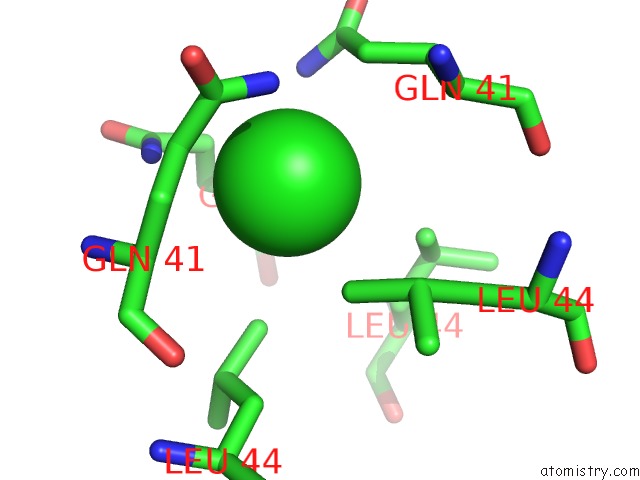
Mono view
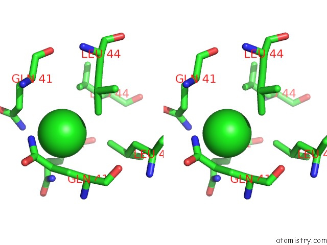
Stereo pair view
Mono view
Stereo pair view
A full contact list of Chlorine with other atoms in the Cl binding
site number 1 of Crystal Structure of the Complex Between GP41 Fragments N36 and C34 Mutant N126K/E137Q within 5.0Å range:
|
Chlorine binding site 2 out of 2 in 3aha
Go back to
Chlorine binding site 2 out
of 2 in the Crystal Structure of the Complex Between GP41 Fragments N36 and C34 Mutant N126K/E137Q
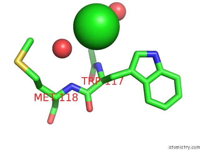
Mono view
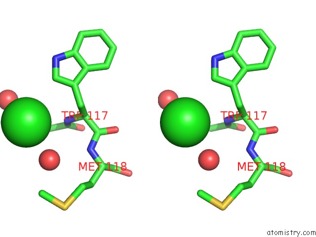
Stereo pair view
Mono view
Stereo pair view
A full contact list of Chlorine with other atoms in the Cl binding
site number 2 of Crystal Structure of the Complex Between GP41 Fragments N36 and C34 Mutant N126K/E137Q within 5.0Å range:
|
Reference:
K.Izumi,
S.Nakamura,
H.Nakano,
K.Shimura,
Y.Sakagami,
S.Oishi,
S.Uchiyama,
T.Ohkubo,
Y.Kobayashi,
N.Fujii,
M.Matsuoka,
E.N.Kodama.
Characterization of Hiv-1 Resistance to A Fusion Inhibitor, N36, Derived From the GP41 Amino Terminal Heptad Repeat. Antiviral Res. 2010.
ISSN: ISSN 0166-3542
PubMed: 20438763
DOI: 10.1016/J.ANTIVIRAL.2010.04.011
Page generated: Sat Jul 20 15:55:25 2024
ISSN: ISSN 0166-3542
PubMed: 20438763
DOI: 10.1016/J.ANTIVIRAL.2010.04.011
Last articles
Zn in 9J0NZn in 9J0O
Zn in 9J0P
Zn in 9FJX
Zn in 9EKB
Zn in 9C0F
Zn in 9CAH
Zn in 9CH0
Zn in 9CH3
Zn in 9CH1