Chlorine »
PDB 3akg-3avh »
3apx »
Chlorine in PDB 3apx: Crystal Structure of the A Variant of Human ALPHA1-Acid Glycoprotein and Chlorpromazine Complex
Protein crystallography data
The structure of Crystal Structure of the A Variant of Human ALPHA1-Acid Glycoprotein and Chlorpromazine Complex, PDB code: 3apx
was solved by
K.Nishi,
T.Ono,
T.Nakamura,
N.Fukunaga,
M.Izumi,
H.Watanabe,
A.Suenaga,
T.Maruyama,
Y.Yamagata,
S.Curry,
M.Otagiri,
with X-Ray Crystallography technique. A brief refinement statistics is given in the table below:
| Resolution Low / High (Å) | 28.88 / 2.20 |
| Space group | P 21 21 21 |
| Cell size a, b, c (Å), α, β, γ (°) | 42.125, 63.077, 64.957, 90.00, 90.00, 90.00 |
| R / Rfree (%) | 20.2 / 26.8 |
Chlorine Binding Sites:
The binding sites of Chlorine atom in the Crystal Structure of the A Variant of Human ALPHA1-Acid Glycoprotein and Chlorpromazine Complex
(pdb code 3apx). This binding sites where shown within
5.0 Angstroms radius around Chlorine atom.
In total only one binding site of Chlorine was determined in the Crystal Structure of the A Variant of Human ALPHA1-Acid Glycoprotein and Chlorpromazine Complex, PDB code: 3apx:
In total only one binding site of Chlorine was determined in the Crystal Structure of the A Variant of Human ALPHA1-Acid Glycoprotein and Chlorpromazine Complex, PDB code: 3apx:
Chlorine binding site 1 out of 1 in 3apx
Go back to
Chlorine binding site 1 out
of 1 in the Crystal Structure of the A Variant of Human ALPHA1-Acid Glycoprotein and Chlorpromazine Complex
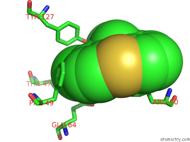
Mono view
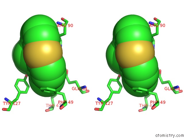
Stereo pair view
Mono view
Stereo pair view
A full contact list of Chlorine with other atoms in the Cl binding
site number 1 of Crystal Structure of the A Variant of Human ALPHA1-Acid Glycoprotein and Chlorpromazine Complex within 5.0Å range:
|
Reference:
K.Nishi,
T.Ono,
T.Nakamura,
N.Fukunaga,
M.Izumi,
H.Watanabe,
A.Suenaga,
T.Maruyama,
Y.Yamagata,
S.Curry,
M.Otagiri.
Structural Insights Into Differences in Drug-Binding Selectivity Between Two Forms of Human ALPHA1-Acid Glycoprotein Genetic Variants, the A and F1*S Forms. J. Biol. Chem. V. 286 14427 2011.
ISSN: ESSN 1083-351X
PubMed: 21349832
DOI: 10.1074/JBC.M110.208926
Page generated: Sat Jul 20 16:01:11 2024
ISSN: ESSN 1083-351X
PubMed: 21349832
DOI: 10.1074/JBC.M110.208926
Last articles
Zn in 9J0NZn in 9J0O
Zn in 9J0P
Zn in 9FJX
Zn in 9EKB
Zn in 9C0F
Zn in 9CAH
Zn in 9CH0
Zn in 9CH3
Zn in 9CH1