Chlorine »
PDB 3bga-3bpr »
3bkd »
Chlorine in PDB 3bkd: High Resolution Crystal Structure of Transmembrane Domain of M2 Protein
Protein crystallography data
The structure of High Resolution Crystal Structure of Transmembrane Domain of M2 Protein, PDB code: 3bkd
was solved by
A.L.Stouffer,
R.Acharya,
D.Salom,
with X-Ray Crystallography technique. A brief refinement statistics is given in the table below:
| Resolution Low / High (Å) | 20.00 / 2.05 |
| Space group | P 1 21 1 |
| Cell size a, b, c (Å), α, β, γ (°) | 38.753, 56.557, 56.009, 90.00, 103.53, 90.00 |
| R / Rfree (%) | 21.9 / 26.9 |
Chlorine Binding Sites:
The binding sites of Chlorine atom in the High Resolution Crystal Structure of Transmembrane Domain of M2 Protein
(pdb code 3bkd). This binding sites where shown within
5.0 Angstroms radius around Chlorine atom.
In total 2 binding sites of Chlorine where determined in the High Resolution Crystal Structure of Transmembrane Domain of M2 Protein, PDB code: 3bkd:
Jump to Chlorine binding site number: 1; 2;
In total 2 binding sites of Chlorine where determined in the High Resolution Crystal Structure of Transmembrane Domain of M2 Protein, PDB code: 3bkd:
Jump to Chlorine binding site number: 1; 2;
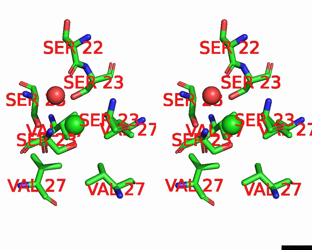
Chlorine binding site 1 out of 2 in 3bkd
Go back to
Chlorine binding site 1 out
of 2 in the High Resolution Crystal Structure of Transmembrane Domain of M2 Protein

Mono view

Stereo pair view
Mono view
Stereo pair view
A full contact list of Chlorine with other atoms in the Cl binding
site number 1 of High Resolution Crystal Structure of Transmembrane Domain of M2 Protein within 5.0Å range:
|
Chlorine binding site 2 out of 2 in 3bkd
Go back to
Chlorine binding site 2 out
of 2 in the High Resolution Crystal Structure of Transmembrane Domain of M2 Protein

Mono view
Stereo pair view
Mono view
Stereo pair view
A full contact list of Chlorine with other atoms in the Cl binding
site number 2 of High Resolution Crystal Structure of Transmembrane Domain of M2 Protein within 5.0Å range:
|
Reference:
A.L.Stouffer,
R.Acharya,
D.Salom,
A.S.Levine,
L.Di Costanzo,
C.S.Soto,
V.Tereshko,
V.Nanda,
S.Stayrook,
W.F.Degrado.
Structural Basis For the Function and Inhibition of An Influenza Virus Proton Channel Nature V. 451 596 2008.
ISSN: ISSN 0028-0836
PubMed: 18235504
DOI: 10.1038/NATURE06528
Page generated: Sat Jul 20 16:35:00 2024
ISSN: ISSN 0028-0836
PubMed: 18235504
DOI: 10.1038/NATURE06528
Last articles
Zn in 9J0NZn in 9J0O
Zn in 9J0P
Zn in 9FJX
Zn in 9EKB
Zn in 9C0F
Zn in 9CAH
Zn in 9CH0
Zn in 9CH3
Zn in 9CH1