Chlorine »
PDB 3bpr-3c4c »
3c44 »
Chlorine in PDB 3c44: Crystal Structure of Hiv-1 Subtype F Dis Extended Duplex Rna Bound to Paromomycin
Protein crystallography data
The structure of Crystal Structure of Hiv-1 Subtype F Dis Extended Duplex Rna Bound to Paromomycin, PDB code: 3c44
was solved by
S.Freisz,
E.Ennifar,
P.Dumas,
with X-Ray Crystallography technique. A brief refinement statistics is given in the table below:
| Resolution Low / High (Å) | 26.55 / 2.00 |
| Space group | P 21 21 2 |
| Cell size a, b, c (Å), α, β, γ (°) | 30.818, 95.703, 47.856, 90.00, 90.00, 90.00 |
| R / Rfree (%) | 22.4 / 24.7 |
Other elements in 3c44:
The structure of Crystal Structure of Hiv-1 Subtype F Dis Extended Duplex Rna Bound to Paromomycin also contains other interesting chemical elements:
| Potassium | (K) | 4 atoms |
Chlorine Binding Sites:
The binding sites of Chlorine atom in the Crystal Structure of Hiv-1 Subtype F Dis Extended Duplex Rna Bound to Paromomycin
(pdb code 3c44). This binding sites where shown within
5.0 Angstroms radius around Chlorine atom.
In total 2 binding sites of Chlorine where determined in the Crystal Structure of Hiv-1 Subtype F Dis Extended Duplex Rna Bound to Paromomycin, PDB code: 3c44:
Jump to Chlorine binding site number: 1; 2;
In total 2 binding sites of Chlorine where determined in the Crystal Structure of Hiv-1 Subtype F Dis Extended Duplex Rna Bound to Paromomycin, PDB code: 3c44:
Jump to Chlorine binding site number: 1; 2;
Chlorine binding site 1 out of 2 in 3c44
Go back to
Chlorine binding site 1 out
of 2 in the Crystal Structure of Hiv-1 Subtype F Dis Extended Duplex Rna Bound to Paromomycin
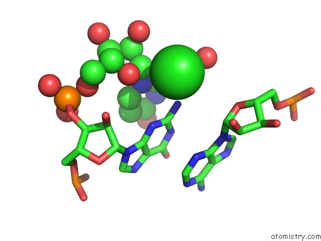
Mono view
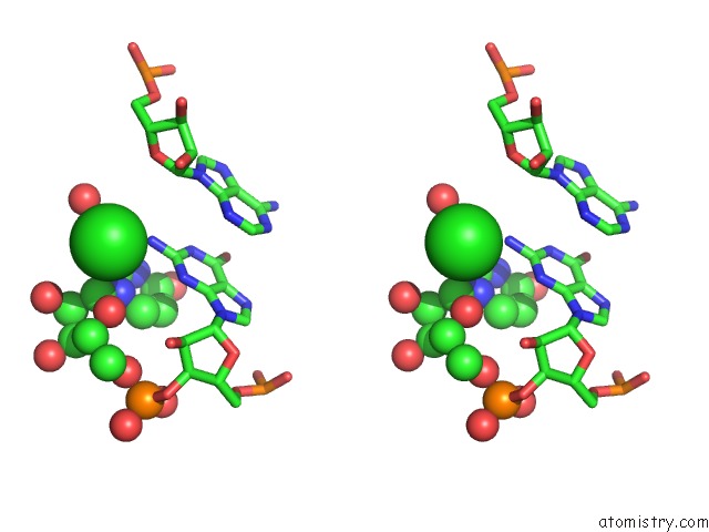
Stereo pair view
Mono view
Stereo pair view
A full contact list of Chlorine with other atoms in the Cl binding
site number 1 of Crystal Structure of Hiv-1 Subtype F Dis Extended Duplex Rna Bound to Paromomycin within 5.0Å range:
|
Chlorine binding site 2 out of 2 in 3c44
Go back to
Chlorine binding site 2 out
of 2 in the Crystal Structure of Hiv-1 Subtype F Dis Extended Duplex Rna Bound to Paromomycin
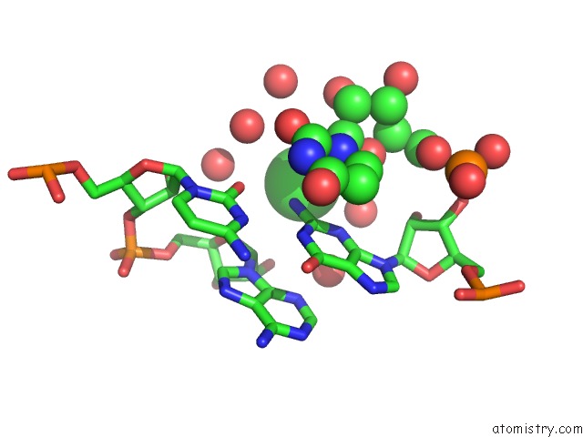
Mono view
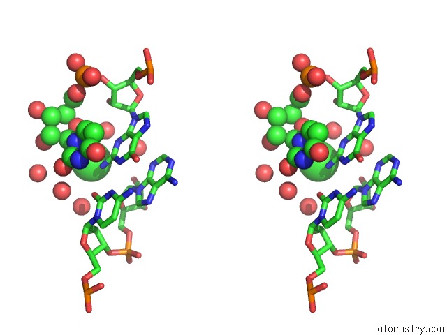
Stereo pair view
Mono view
Stereo pair view
A full contact list of Chlorine with other atoms in the Cl binding
site number 2 of Crystal Structure of Hiv-1 Subtype F Dis Extended Duplex Rna Bound to Paromomycin within 5.0Å range:
|
Reference:
S.Freisz,
K.Lang,
R.Micura,
P.Dumas,
E.Ennifar.
Binding of Aminoglycoside Antibiotics to the Duplex Form of the Hiv-1 Genomic Rna Dimerization Initiation Site. Angew.Chem.Int.Ed.Engl. V. 47 4110 2008.
ISSN: ISSN 0570-0833
PubMed: 18435520
DOI: 10.1002/ANIE.200800726
Page generated: Sat Jul 20 16:49:54 2024
ISSN: ISSN 0570-0833
PubMed: 18435520
DOI: 10.1002/ANIE.200800726
Last articles
Zn in 9JYWZn in 9IR4
Zn in 9IR3
Zn in 9GMX
Zn in 9GMW
Zn in 9JEJ
Zn in 9ERF
Zn in 9ERE
Zn in 9EGV
Zn in 9EGW