Chlorine »
PDB 3bqc-3c4w »
3c4w »
Chlorine in PDB 3c4w: Crystal Structure of G Protein Coupled Receptor Kinase 1 Bound to Atp and Magnesium Chloride at 2.7A
Enzymatic activity of Crystal Structure of G Protein Coupled Receptor Kinase 1 Bound to Atp and Magnesium Chloride at 2.7A
All present enzymatic activity of Crystal Structure of G Protein Coupled Receptor Kinase 1 Bound to Atp and Magnesium Chloride at 2.7A:
2.7.11.14;
2.7.11.14;
Protein crystallography data
The structure of Crystal Structure of G Protein Coupled Receptor Kinase 1 Bound to Atp and Magnesium Chloride at 2.7A, PDB code: 3c4w
was solved by
P.Singh,
J.J.G.Tesmer,
with X-Ray Crystallography technique. A brief refinement statistics is given in the table below:
| Resolution Low / High (Å) | 19.98 / 2.70 |
| Space group | C 1 2 1 |
| Cell size a, b, c (Å), α, β, γ (°) | 202.889, 55.054, 122.699, 90.00, 100.82, 90.00 |
| R / Rfree (%) | 19.2 / n/a |
Other elements in 3c4w:
The structure of Crystal Structure of G Protein Coupled Receptor Kinase 1 Bound to Atp and Magnesium Chloride at 2.7A also contains other interesting chemical elements:
| Magnesium | (Mg) | 3 atoms |
Chlorine Binding Sites:
The binding sites of Chlorine atom in the Crystal Structure of G Protein Coupled Receptor Kinase 1 Bound to Atp and Magnesium Chloride at 2.7A
(pdb code 3c4w). This binding sites where shown within
5.0 Angstroms radius around Chlorine atom.
In total 3 binding sites of Chlorine where determined in the Crystal Structure of G Protein Coupled Receptor Kinase 1 Bound to Atp and Magnesium Chloride at 2.7A, PDB code: 3c4w:
Jump to Chlorine binding site number: 1; 2; 3;
In total 3 binding sites of Chlorine where determined in the Crystal Structure of G Protein Coupled Receptor Kinase 1 Bound to Atp and Magnesium Chloride at 2.7A, PDB code: 3c4w:
Jump to Chlorine binding site number: 1; 2; 3;
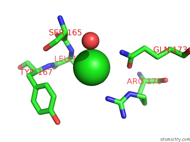
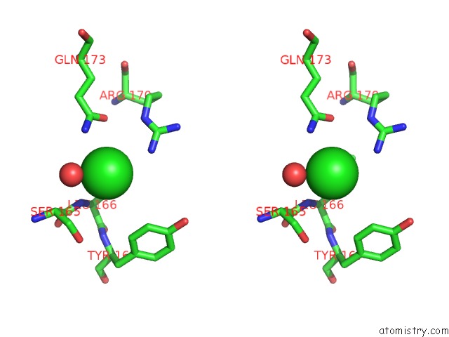
Chlorine binding site 1 out of 3 in 3c4w
Go back to
Chlorine binding site 1 out
of 3 in the Crystal Structure of G Protein Coupled Receptor Kinase 1 Bound to Atp and Magnesium Chloride at 2.7A
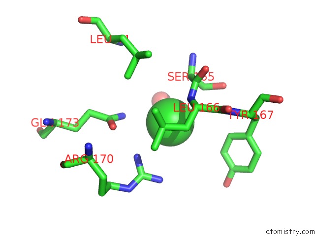
Mono view
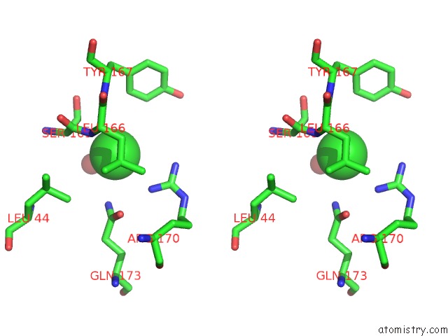
Stereo pair view
Mono view
Stereo pair view
A full contact list of Chlorine with other atoms in the Cl binding
site number 1 of Crystal Structure of G Protein Coupled Receptor Kinase 1 Bound to Atp and Magnesium Chloride at 2.7A within 5.0Å range:
|
Chlorine binding site 2 out of 3 in 3c4w
Go back to
Chlorine binding site 2 out
of 3 in the Crystal Structure of G Protein Coupled Receptor Kinase 1 Bound to Atp and Magnesium Chloride at 2.7A
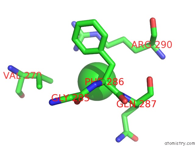
Mono view
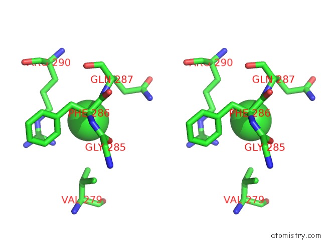
Stereo pair view
Mono view
Stereo pair view
A full contact list of Chlorine with other atoms in the Cl binding
site number 2 of Crystal Structure of G Protein Coupled Receptor Kinase 1 Bound to Atp and Magnesium Chloride at 2.7A within 5.0Å range:
|
Chlorine binding site 3 out of 3 in 3c4w
Go back to
Chlorine binding site 3 out
of 3 in the Crystal Structure of G Protein Coupled Receptor Kinase 1 Bound to Atp and Magnesium Chloride at 2.7A
Mono view
Stereo pair view
Mono view
Stereo pair view
A full contact list of Chlorine with other atoms in the Cl binding
site number 3 of Crystal Structure of G Protein Coupled Receptor Kinase 1 Bound to Atp and Magnesium Chloride at 2.7A within 5.0Å range:
|
Reference:
P.Singh,
B.Wang,
T.Maeda,
K.Palczewski,
J.J.Tesmer.
Structures of Rhodopsin Kinase in Different Ligand States Reveal Key Elements Involved in G Protein-Coupled Receptor Kinase Activation. J.Biol.Chem. V. 283 14053 2008.
ISSN: ISSN 0021-9258
PubMed: 18339619
DOI: 10.1074/JBC.M708974200
Page generated: Fri Jul 11 03:36:54 2025
ISSN: ISSN 0021-9258
PubMed: 18339619
DOI: 10.1074/JBC.M708974200
Last articles
Cl in 5TM7Cl in 5TM4
Cl in 5TM3
Cl in 5TL4
Cl in 5TM2
Cl in 5TLL
Cl in 5TLS
Cl in 5TKS
Cl in 5TKU
Cl in 5TKT