Chlorine »
PDB 3ccq-3ck1 »
3cd3 »
Chlorine in PDB 3cd3: Crystal Structure of Phosphorylated Human Feline Sarcoma Viral Oncogene Homologue (V-Fes) in Complex with Staurosporine and A Consensus Peptide
Enzymatic activity of Crystal Structure of Phosphorylated Human Feline Sarcoma Viral Oncogene Homologue (V-Fes) in Complex with Staurosporine and A Consensus Peptide
All present enzymatic activity of Crystal Structure of Phosphorylated Human Feline Sarcoma Viral Oncogene Homologue (V-Fes) in Complex with Staurosporine and A Consensus Peptide:
2.7.10.2;
2.7.10.2;
Protein crystallography data
The structure of Crystal Structure of Phosphorylated Human Feline Sarcoma Viral Oncogene Homologue (V-Fes) in Complex with Staurosporine and A Consensus Peptide, PDB code: 3cd3
was solved by
P.Filippakopoulos,
E.Salah,
C.Cooper,
S.S.Picaud,
J.M.Elkins,
F.Von Delft,
C.H.Arrowsmith,
A.M.Edwards,
J.Weigelt,
C.Bountra,
S.Knapp,
Structuralgenomics Consortium (Sgc),
with X-Ray Crystallography technique. A brief refinement statistics is given in the table below:
| Resolution Low / High (Å) | 15.35 / 1.98 |
| Space group | P 21 21 21 |
| Cell size a, b, c (Å), α, β, γ (°) | 35.451, 76.905, 150.632, 90.00, 90.00, 90.00 |
| R / Rfree (%) | 18.5 / 24.7 |
Chlorine Binding Sites:
The binding sites of Chlorine atom in the Crystal Structure of Phosphorylated Human Feline Sarcoma Viral Oncogene Homologue (V-Fes) in Complex with Staurosporine and A Consensus Peptide
(pdb code 3cd3). This binding sites where shown within
5.0 Angstroms radius around Chlorine atom.
In total 2 binding sites of Chlorine where determined in the Crystal Structure of Phosphorylated Human Feline Sarcoma Viral Oncogene Homologue (V-Fes) in Complex with Staurosporine and A Consensus Peptide, PDB code: 3cd3:
Jump to Chlorine binding site number: 1; 2;
In total 2 binding sites of Chlorine where determined in the Crystal Structure of Phosphorylated Human Feline Sarcoma Viral Oncogene Homologue (V-Fes) in Complex with Staurosporine and A Consensus Peptide, PDB code: 3cd3:
Jump to Chlorine binding site number: 1; 2;
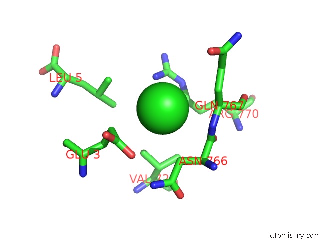
Chlorine binding site 1 out of 2 in 3cd3
Go back to
Chlorine binding site 1 out
of 2 in the Crystal Structure of Phosphorylated Human Feline Sarcoma Viral Oncogene Homologue (V-Fes) in Complex with Staurosporine and A Consensus Peptide
Mono view
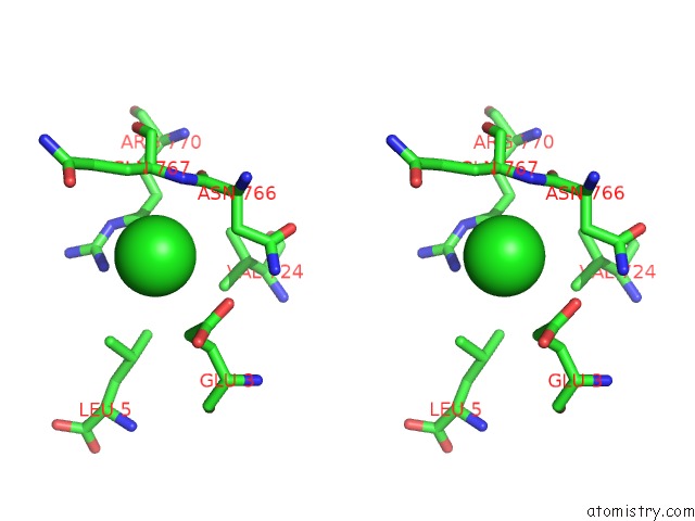
Stereo pair view
Mono view
Stereo pair view
A full contact list of Chlorine with other atoms in the Cl binding
site number 1 of Crystal Structure of Phosphorylated Human Feline Sarcoma Viral Oncogene Homologue (V-Fes) in Complex with Staurosporine and A Consensus Peptide within 5.0Å range:
|
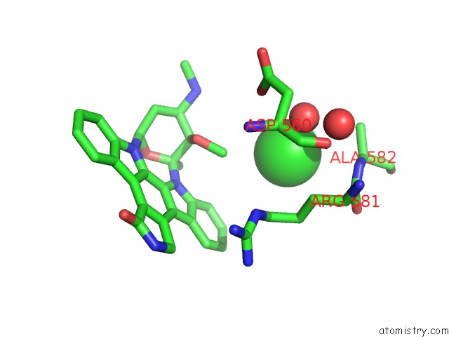
Chlorine binding site 2 out of 2 in 3cd3
Go back to
Chlorine binding site 2 out
of 2 in the Crystal Structure of Phosphorylated Human Feline Sarcoma Viral Oncogene Homologue (V-Fes) in Complex with Staurosporine and A Consensus Peptide
Mono view
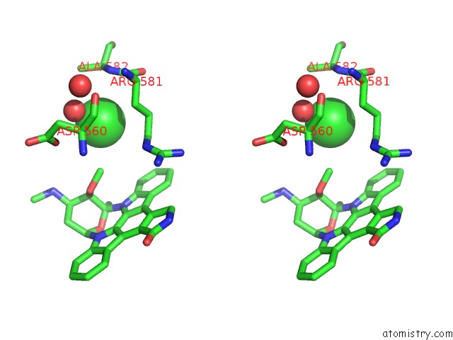
Stereo pair view
Mono view
Stereo pair view
A full contact list of Chlorine with other atoms in the Cl binding
site number 2 of Crystal Structure of Phosphorylated Human Feline Sarcoma Viral Oncogene Homologue (V-Fes) in Complex with Staurosporine and A Consensus Peptide within 5.0Å range:
|
Reference:
P.Filippakopoulos,
M.Kofler,
O.Hantschel,
G.D.Gish,
F.Grebien,
E.Salah,
P.Neudecker,
L.E.Kay,
B.E.Turk,
G.Superti-Furga,
T.Pawson,
S.Knapp.
Structural Coupling of SH2-Kinase Domains Links Fes and Abl Substrate Recognition and Kinase Activation Cell(Cambridge,Mass.) V. 134 793 2008.
ISSN: ISSN 0092-8674
PubMed: 18775312
DOI: 10.1016/J.CELL.2008.07.047
Page generated: Sat Jul 20 17:19:57 2024
ISSN: ISSN 0092-8674
PubMed: 18775312
DOI: 10.1016/J.CELL.2008.07.047
Last articles
Zn in 9J0NZn in 9J0O
Zn in 9J0P
Zn in 9FJX
Zn in 9EKB
Zn in 9C0F
Zn in 9CAH
Zn in 9CH0
Zn in 9CH3
Zn in 9CH1