Chlorine »
PDB 3ct9-3d44 »
3cxp »
Chlorine in PDB 3cxp: Crystal Structure of Human Glucosamine 6-Phosphate N- Acetyltransferase 1 Mutant E156A
Enzymatic activity of Crystal Structure of Human Glucosamine 6-Phosphate N- Acetyltransferase 1 Mutant E156A
All present enzymatic activity of Crystal Structure of Human Glucosamine 6-Phosphate N- Acetyltransferase 1 Mutant E156A:
2.3.1.4;
2.3.1.4;
Protein crystallography data
The structure of Crystal Structure of Human Glucosamine 6-Phosphate N- Acetyltransferase 1 Mutant E156A, PDB code: 3cxp
was solved by
J.Wang,
X.Liu,
L.-F.Li,
X.-D.Su,
with X-Ray Crystallography technique. A brief refinement statistics is given in the table below:
| Resolution Low / High (Å) | 19.30 / 2.01 |
| Space group | P 43 21 2 |
| Cell size a, b, c (Å), α, β, γ (°) | 51.340, 51.340, 142.610, 90.00, 90.00, 90.00 |
| R / Rfree (%) | 19.9 / 23.2 |
Chlorine Binding Sites:
The binding sites of Chlorine atom in the Crystal Structure of Human Glucosamine 6-Phosphate N- Acetyltransferase 1 Mutant E156A
(pdb code 3cxp). This binding sites where shown within
5.0 Angstroms radius around Chlorine atom.
In total only one binding site of Chlorine was determined in the Crystal Structure of Human Glucosamine 6-Phosphate N- Acetyltransferase 1 Mutant E156A, PDB code: 3cxp:
In total only one binding site of Chlorine was determined in the Crystal Structure of Human Glucosamine 6-Phosphate N- Acetyltransferase 1 Mutant E156A, PDB code: 3cxp:
Chlorine binding site 1 out of 1 in 3cxp
Go back to
Chlorine binding site 1 out
of 1 in the Crystal Structure of Human Glucosamine 6-Phosphate N- Acetyltransferase 1 Mutant E156A
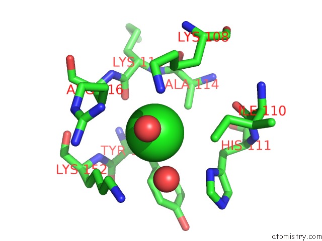
Mono view
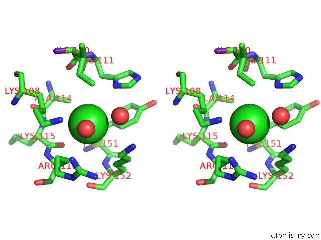
Stereo pair view
Mono view
Stereo pair view
A full contact list of Chlorine with other atoms in the Cl binding
site number 1 of Crystal Structure of Human Glucosamine 6-Phosphate N- Acetyltransferase 1 Mutant E156A within 5.0Å range:
|
Reference:
J.Wang,
X.Liu,
Y.-H.Liang,
L.-F.Li,
X.-D.Su.
Acceptor Substrate Binding Revealed By Crystal Structure of Human Glucosamine-6-Phosphate N-Acetyltransferase 1 Febs Lett. V. 582 2973 2008.
ISSN: ISSN 0014-5793
PubMed: 18675810
DOI: 10.1016/J.FEBSLET.2008.07.040
Page generated: Sat Jul 20 17:52:22 2024
ISSN: ISSN 0014-5793
PubMed: 18675810
DOI: 10.1016/J.FEBSLET.2008.07.040
Last articles
Zn in 9J0NZn in 9J0O
Zn in 9J0P
Zn in 9FJX
Zn in 9EKB
Zn in 9C0F
Zn in 9CAH
Zn in 9CH0
Zn in 9CH3
Zn in 9CH1