Chlorine »
PDB 3d4i-3dgo »
3da2 »
Chlorine in PDB 3da2: X-Ray Structure of Human Carbonic Anhydrase 13 in Complex with Inhibitor
Enzymatic activity of X-Ray Structure of Human Carbonic Anhydrase 13 in Complex with Inhibitor
All present enzymatic activity of X-Ray Structure of Human Carbonic Anhydrase 13 in Complex with Inhibitor:
4.2.1.1;
4.2.1.1;
Protein crystallography data
The structure of X-Ray Structure of Human Carbonic Anhydrase 13 in Complex with Inhibitor, PDB code: 3da2
was solved by
E.S.Pilka,
S.S.Picaud,
W.W.Yue,
O.N.F.King,
J.E.Bray,
P.Filippakopoulos,
A.K.Roos,
A.C.W.Pike,
F.Von Delft,
C.H.Arrowsmith,
M.Wikstrom,
A.M.Edwards,
C.Bountra,
U.Oppermann,
Structural Genomics Consortium(Sgc),
with X-Ray Crystallography technique. A brief refinement statistics is given in the table below:
| Resolution Low / High (Å) | 43.00 / 2.05 |
| Space group | C 1 2 1 |
| Cell size a, b, c (Å), α, β, γ (°) | 153.637, 45.138, 87.938, 90.00, 116.28, 90.00 |
| R / Rfree (%) | 15.9 / 22.1 |
Other elements in 3da2:
The structure of X-Ray Structure of Human Carbonic Anhydrase 13 in Complex with Inhibitor also contains other interesting chemical elements:
| Zinc | (Zn) | 2 atoms |
Chlorine Binding Sites:
The binding sites of Chlorine atom in the X-Ray Structure of Human Carbonic Anhydrase 13 in Complex with Inhibitor
(pdb code 3da2). This binding sites where shown within
5.0 Angstroms radius around Chlorine atom.
In total 4 binding sites of Chlorine where determined in the X-Ray Structure of Human Carbonic Anhydrase 13 in Complex with Inhibitor, PDB code: 3da2:
Jump to Chlorine binding site number: 1; 2; 3; 4;
In total 4 binding sites of Chlorine where determined in the X-Ray Structure of Human Carbonic Anhydrase 13 in Complex with Inhibitor, PDB code: 3da2:
Jump to Chlorine binding site number: 1; 2; 3; 4;
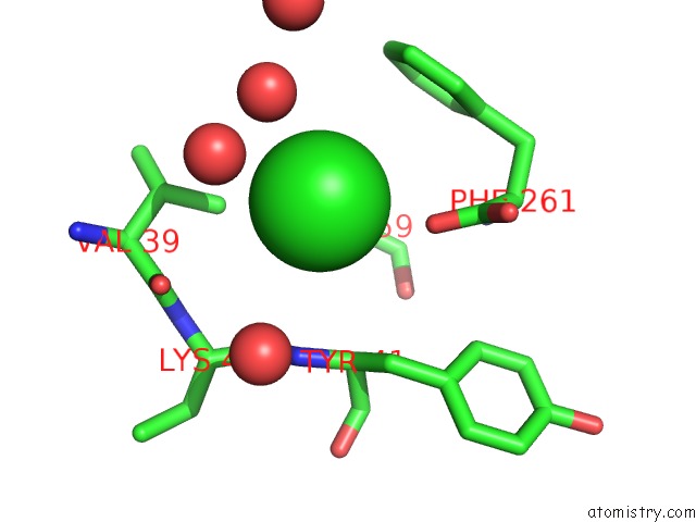
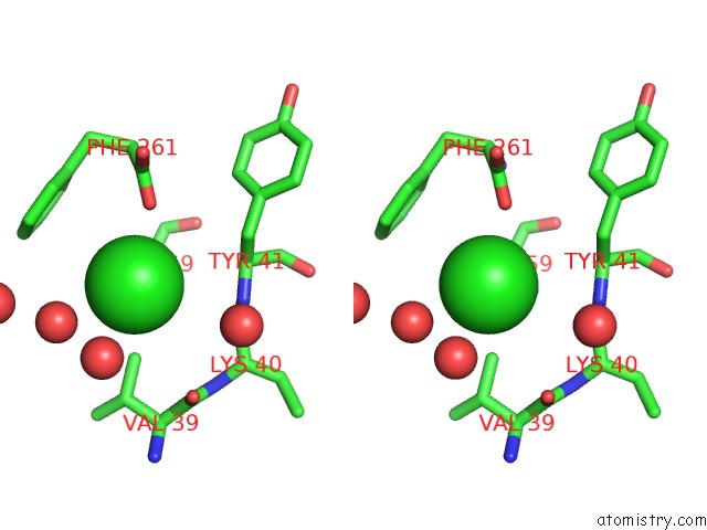
Chlorine binding site 1 out of 4 in 3da2
Go back to
Chlorine binding site 1 out
of 4 in the X-Ray Structure of Human Carbonic Anhydrase 13 in Complex with Inhibitor
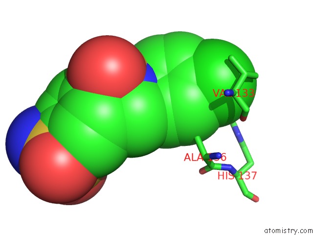
Mono view
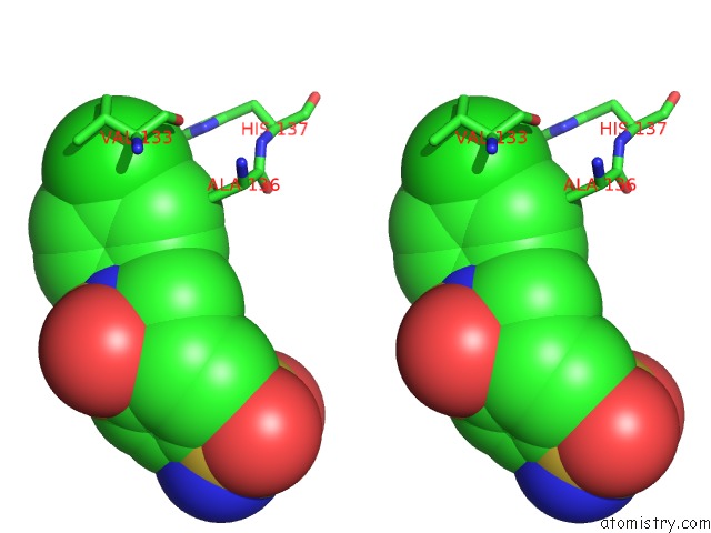
Stereo pair view
Mono view
Stereo pair view
A full contact list of Chlorine with other atoms in the Cl binding
site number 1 of X-Ray Structure of Human Carbonic Anhydrase 13 in Complex with Inhibitor within 5.0Å range:
|
Chlorine binding site 2 out of 4 in 3da2
Go back to
Chlorine binding site 2 out
of 4 in the X-Ray Structure of Human Carbonic Anhydrase 13 in Complex with Inhibitor
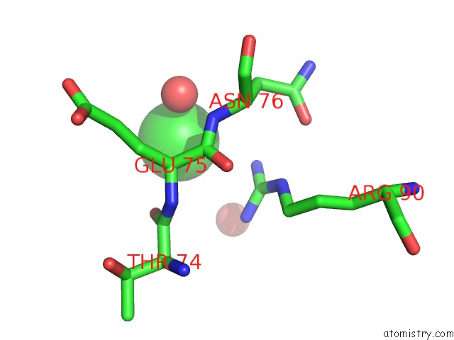
Mono view
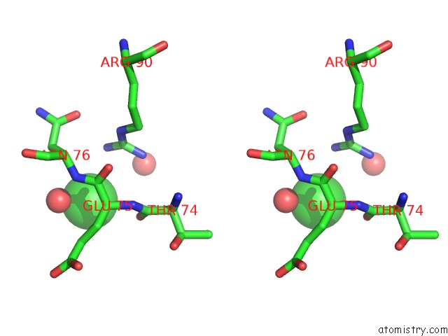
Stereo pair view
Mono view
Stereo pair view
A full contact list of Chlorine with other atoms in the Cl binding
site number 2 of X-Ray Structure of Human Carbonic Anhydrase 13 in Complex with Inhibitor within 5.0Å range:
|
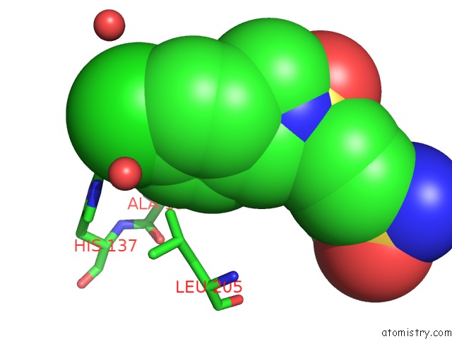
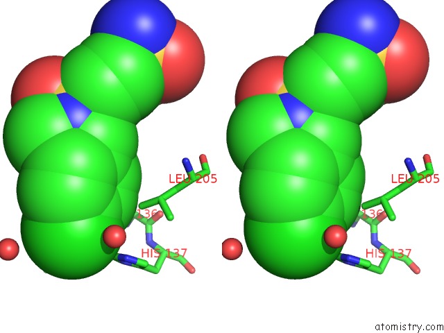
Chlorine binding site 3 out of 4 in 3da2
Go back to
Chlorine binding site 3 out
of 4 in the X-Ray Structure of Human Carbonic Anhydrase 13 in Complex with Inhibitor
Mono view
Stereo pair view
Mono view
Stereo pair view
A full contact list of Chlorine with other atoms in the Cl binding
site number 3 of X-Ray Structure of Human Carbonic Anhydrase 13 in Complex with Inhibitor within 5.0Å range:
|
Chlorine binding site 4 out of 4 in 3da2
Go back to
Chlorine binding site 4 out
of 4 in the X-Ray Structure of Human Carbonic Anhydrase 13 in Complex with Inhibitor
Mono view
Stereo pair view
Mono view
Stereo pair view
A full contact list of Chlorine with other atoms in the Cl binding
site number 4 of X-Ray Structure of Human Carbonic Anhydrase 13 in Complex with Inhibitor within 5.0Å range:
|
Reference:
E.S.Pilka,
S.S.Picaud,
W.W.Yue,
O.N.F.King,
J.E.Bray,
P.Filippakopoulos,
A.K.Roos,
A.C.W.Pike,
F.Von Delft,
C.H.Arrowsmith,
M.Wikstrom,
A.M.Edwards,
C.Bountra,
U.Oppermann.
X-Ray Structure of Human Carbonic Anhydrase 13 in Complex with Inhibitor. To Be Published.
Page generated: Sat Jul 20 18:05:57 2024
Last articles
Zn in 9J0NZn in 9J0O
Zn in 9J0P
Zn in 9FJX
Zn in 9EKB
Zn in 9C0F
Zn in 9CAH
Zn in 9CH0
Zn in 9CH3
Zn in 9CH1