Chlorine »
PDB 3d4i-3dgo »
3daq »
Chlorine in PDB 3daq: Crystal Structure of Dihydrodipicolinate Synthase From Methicillin- Resistant Staphylococcus Aureus
Enzymatic activity of Crystal Structure of Dihydrodipicolinate Synthase From Methicillin- Resistant Staphylococcus Aureus
All present enzymatic activity of Crystal Structure of Dihydrodipicolinate Synthase From Methicillin- Resistant Staphylococcus Aureus:
4.2.1.52;
4.2.1.52;
Protein crystallography data
The structure of Crystal Structure of Dihydrodipicolinate Synthase From Methicillin- Resistant Staphylococcus Aureus, PDB code: 3daq
was solved by
R.C.J.Dobson,
B.R.Burgess,
G.B.Jameson,
J.A.Gerrard,
M.W.Parker,
M.A.Perugini,
with X-Ray Crystallography technique. A brief refinement statistics is given in the table below:
| Resolution Low / High (Å) | 30.25 / 1.45 |
| Space group | P 1 |
| Cell size a, b, c (Å), α, β, γ (°) | 65.410, 67.573, 77.998, 90.13, 68.85, 72.29 |
| R / Rfree (%) | 13.2 / 16.1 |
Chlorine Binding Sites:
The binding sites of Chlorine atom in the Crystal Structure of Dihydrodipicolinate Synthase From Methicillin- Resistant Staphylococcus Aureus
(pdb code 3daq). This binding sites where shown within
5.0 Angstroms radius around Chlorine atom.
In total 4 binding sites of Chlorine where determined in the Crystal Structure of Dihydrodipicolinate Synthase From Methicillin- Resistant Staphylococcus Aureus, PDB code: 3daq:
Jump to Chlorine binding site number: 1; 2; 3; 4;
In total 4 binding sites of Chlorine where determined in the Crystal Structure of Dihydrodipicolinate Synthase From Methicillin- Resistant Staphylococcus Aureus, PDB code: 3daq:
Jump to Chlorine binding site number: 1; 2; 3; 4;
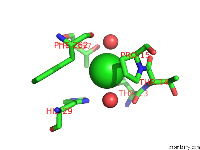
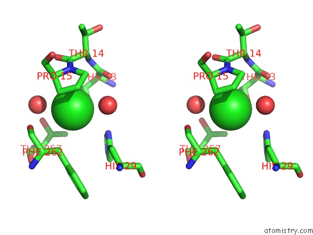
Chlorine binding site 1 out of 4 in 3daq
Go back to
Chlorine binding site 1 out
of 4 in the Crystal Structure of Dihydrodipicolinate Synthase From Methicillin- Resistant Staphylococcus Aureus
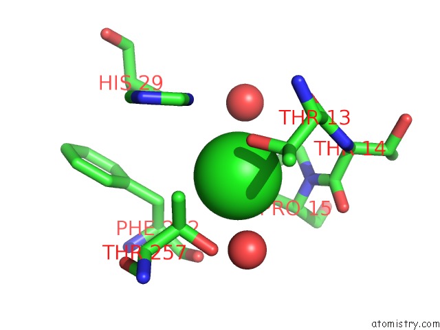
Mono view
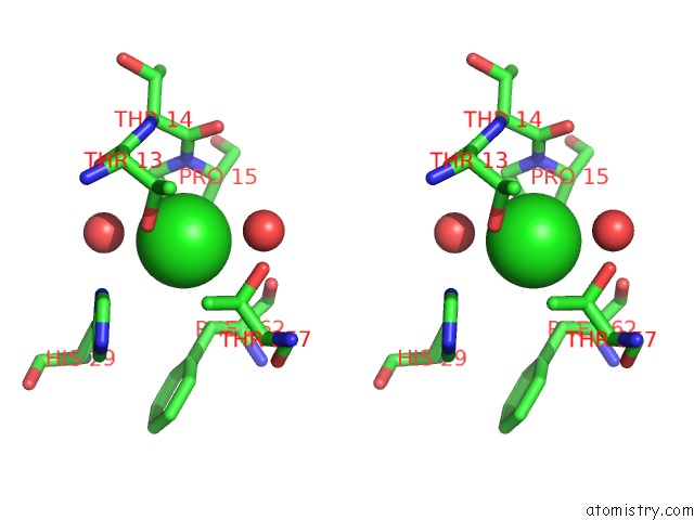
Stereo pair view
Mono view
Stereo pair view
A full contact list of Chlorine with other atoms in the Cl binding
site number 1 of Crystal Structure of Dihydrodipicolinate Synthase From Methicillin- Resistant Staphylococcus Aureus within 5.0Å range:
|
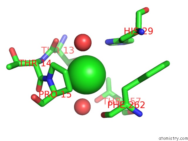
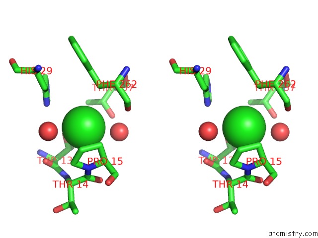
Chlorine binding site 2 out of 4 in 3daq
Go back to
Chlorine binding site 2 out
of 4 in the Crystal Structure of Dihydrodipicolinate Synthase From Methicillin- Resistant Staphylococcus Aureus
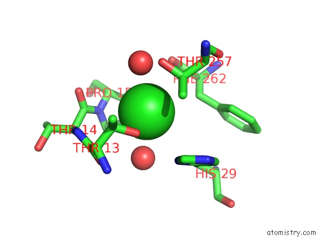
Mono view
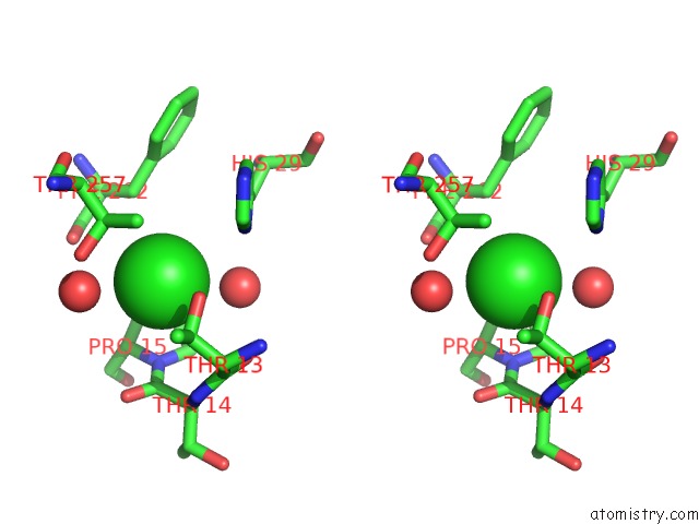
Stereo pair view
Mono view
Stereo pair view
A full contact list of Chlorine with other atoms in the Cl binding
site number 2 of Crystal Structure of Dihydrodipicolinate Synthase From Methicillin- Resistant Staphylococcus Aureus within 5.0Å range:
|
Chlorine binding site 3 out of 4 in 3daq
Go back to
Chlorine binding site 3 out
of 4 in the Crystal Structure of Dihydrodipicolinate Synthase From Methicillin- Resistant Staphylococcus Aureus
Mono view
Stereo pair view
Mono view
Stereo pair view
A full contact list of Chlorine with other atoms in the Cl binding
site number 3 of Crystal Structure of Dihydrodipicolinate Synthase From Methicillin- Resistant Staphylococcus Aureus within 5.0Å range:
|
Chlorine binding site 4 out of 4 in 3daq
Go back to
Chlorine binding site 4 out
of 4 in the Crystal Structure of Dihydrodipicolinate Synthase From Methicillin- Resistant Staphylococcus Aureus
Mono view
Stereo pair view
Mono view
Stereo pair view
A full contact list of Chlorine with other atoms in the Cl binding
site number 4 of Crystal Structure of Dihydrodipicolinate Synthase From Methicillin- Resistant Staphylococcus Aureus within 5.0Å range:
|
Reference:
B.R.Burgess,
R.C.J.Dobson,
M.F.Bailey,
S.C.Atkinson,
M.D.W.Griffin,
G.B.Jameson,
M.W.Parker,
J.A.Gerrard,
M.A.Perugini.
Structure and Evolution of A Novel Dimeric Enzyme From A Clinically-Important Bacterial Pathogen. J.Biol.Chem. 2008.
ISSN: ESSN 1083-351X
PubMed: 18684709
DOI: 10.1074/JBC.M804231200
Page generated: Fri Jul 11 04:23:47 2025
ISSN: ESSN 1083-351X
PubMed: 18684709
DOI: 10.1074/JBC.M804231200
Last articles
Cl in 3UUBCl in 3UUE
Cl in 3UU8
Cl in 3UU6
Cl in 3UU5
Cl in 3UTA
Cl in 3USC
Cl in 3UTU
Cl in 3USE
Cl in 3UT9