Chlorine »
PDB 3dgq-3dn8 »
3dkx »
Chlorine in PDB 3dkx: Crystal Structure of the Replication Initiator Protein Encoded on Plasmid PMV158 (Repb), Trigonal Form, to 2.7 Ang Resolution
Protein crystallography data
The structure of Crystal Structure of the Replication Initiator Protein Encoded on Plasmid PMV158 (Repb), Trigonal Form, to 2.7 Ang Resolution, PDB code: 3dkx
was solved by
D.R.Boer,
J.A.Ruiz-Maso,
A.G.Blanco,
M.Vives-Llacer,
I.Uson,
F.X.Gomis-Ruth,
M.Espinosa,
G.Del Solar,
M.Coll,
with X-Ray Crystallography technique. A brief refinement statistics is given in the table below:
| Resolution Low / High (Å) | 24.95 / 2.70 |
| Space group | P 32 2 1 |
| Cell size a, b, c (Å), α, β, γ (°) | 85.611, 85.611, 245.530, 90.00, 90.00, 120.00 |
| R / Rfree (%) | 22.8 / 27.6 |
Other elements in 3dkx:
The structure of Crystal Structure of the Replication Initiator Protein Encoded on Plasmid PMV158 (Repb), Trigonal Form, to 2.7 Ang Resolution also contains other interesting chemical elements:
| Magnesium | (Mg) | 1 atom |
| Manganese | (Mn) | 3 atoms |
Chlorine Binding Sites:
The binding sites of Chlorine atom in the Crystal Structure of the Replication Initiator Protein Encoded on Plasmid PMV158 (Repb), Trigonal Form, to 2.7 Ang Resolution
(pdb code 3dkx). This binding sites where shown within
5.0 Angstroms radius around Chlorine atom.
In total 3 binding sites of Chlorine where determined in the Crystal Structure of the Replication Initiator Protein Encoded on Plasmid PMV158 (Repb), Trigonal Form, to 2.7 Ang Resolution, PDB code: 3dkx:
Jump to Chlorine binding site number: 1; 2; 3;
In total 3 binding sites of Chlorine where determined in the Crystal Structure of the Replication Initiator Protein Encoded on Plasmid PMV158 (Repb), Trigonal Form, to 2.7 Ang Resolution, PDB code: 3dkx:
Jump to Chlorine binding site number: 1; 2; 3;
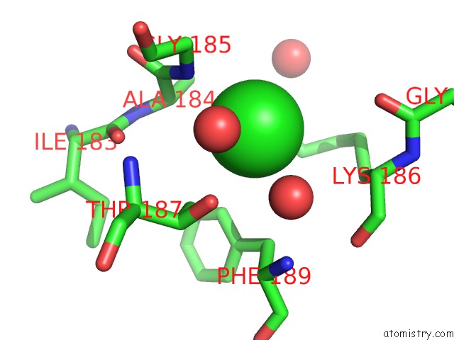
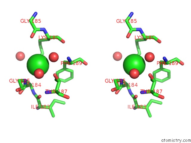
Chlorine binding site 1 out of 3 in 3dkx
Go back to
Chlorine binding site 1 out
of 3 in the Crystal Structure of the Replication Initiator Protein Encoded on Plasmid PMV158 (Repb), Trigonal Form, to 2.7 Ang Resolution
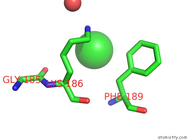
Mono view
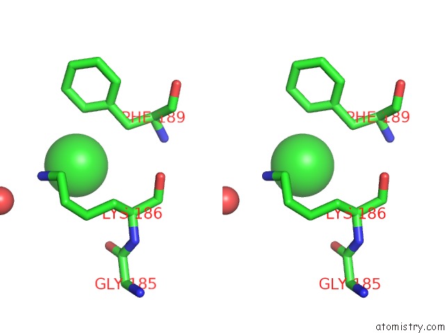
Stereo pair view
Mono view
Stereo pair view
A full contact list of Chlorine with other atoms in the Cl binding
site number 1 of Crystal Structure of the Replication Initiator Protein Encoded on Plasmid PMV158 (Repb), Trigonal Form, to 2.7 Ang Resolution within 5.0Å range:
|
Chlorine binding site 2 out of 3 in 3dkx
Go back to
Chlorine binding site 2 out
of 3 in the Crystal Structure of the Replication Initiator Protein Encoded on Plasmid PMV158 (Repb), Trigonal Form, to 2.7 Ang Resolution
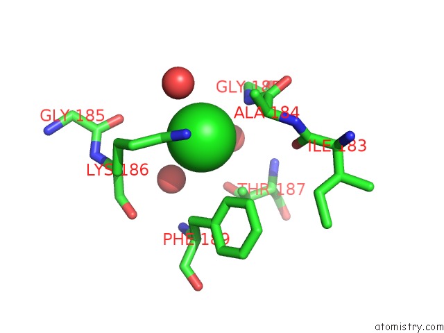
Mono view
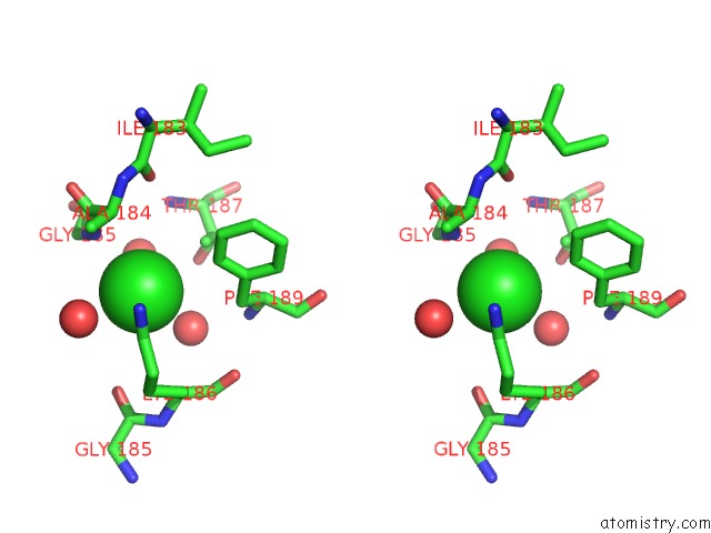
Stereo pair view
Mono view
Stereo pair view
A full contact list of Chlorine with other atoms in the Cl binding
site number 2 of Crystal Structure of the Replication Initiator Protein Encoded on Plasmid PMV158 (Repb), Trigonal Form, to 2.7 Ang Resolution within 5.0Å range:
|
Chlorine binding site 3 out of 3 in 3dkx
Go back to
Chlorine binding site 3 out
of 3 in the Crystal Structure of the Replication Initiator Protein Encoded on Plasmid PMV158 (Repb), Trigonal Form, to 2.7 Ang Resolution
Mono view
Stereo pair view
Mono view
Stereo pair view
A full contact list of Chlorine with other atoms in the Cl binding
site number 3 of Crystal Structure of the Replication Initiator Protein Encoded on Plasmid PMV158 (Repb), Trigonal Form, to 2.7 Ang Resolution within 5.0Å range:
|
Reference:
D.R.Boer,
J.A.Ruiz-Maso,
J.R.Lopez-Blanco,
A.G.Blanco,
M.Vives-Llacer,
P.Chacon,
I.Uson,
F.X.Gomis-Ruth,
M.Espinosa,
O.Llorca,
G.Del Solar,
M.Coll.
Plasmid Replication Initiator Repb Forms A Hexamer Reminiscent of Ring Helicases and Has Mobile Nuclease Domains Embo J. V. 28 1666 2009.
ISSN: ISSN 0261-4189
PubMed: 19440202
DOI: 10.1038/EMBOJ.2009.125
Page generated: Sat Jul 20 18:21:11 2024
ISSN: ISSN 0261-4189
PubMed: 19440202
DOI: 10.1038/EMBOJ.2009.125
Last articles
Zn in 9J0NZn in 9J0O
Zn in 9J0P
Zn in 9FJX
Zn in 9EKB
Zn in 9C0F
Zn in 9CAH
Zn in 9CH0
Zn in 9CH3
Zn in 9CH1