Chlorine »
PDB 3ecg-3es1 »
3egz »
Chlorine in PDB 3egz: Crystal Structure of An in Vitro Evolved Tetracycline Aptamer and Artificial Riboswitch
Protein crystallography data
The structure of Crystal Structure of An in Vitro Evolved Tetracycline Aptamer and Artificial Riboswitch, PDB code: 3egz
was solved by
H.Xiao,
T.E.Edwards,
A.R.Ferre-D'amare,
with X-Ray Crystallography technique. A brief refinement statistics is given in the table below:
| Resolution Low / High (Å) | 29.31 / 2.20 |
| Space group | P 4 21 2 |
| Cell size a, b, c (Å), α, β, γ (°) | 120.835, 120.835, 55.279, 90.00, 90.00, 90.00 |
| R / Rfree (%) | 21.2 / 26 |
Other elements in 3egz:
The structure of Crystal Structure of An in Vitro Evolved Tetracycline Aptamer and Artificial Riboswitch also contains other interesting chemical elements:
| Magnesium | (Mg) | 12 atoms |
Chlorine Binding Sites:
The binding sites of Chlorine atom in the Crystal Structure of An in Vitro Evolved Tetracycline Aptamer and Artificial Riboswitch
(pdb code 3egz). This binding sites where shown within
5.0 Angstroms radius around Chlorine atom.
In total only one binding site of Chlorine was determined in the Crystal Structure of An in Vitro Evolved Tetracycline Aptamer and Artificial Riboswitch, PDB code: 3egz:
In total only one binding site of Chlorine was determined in the Crystal Structure of An in Vitro Evolved Tetracycline Aptamer and Artificial Riboswitch, PDB code: 3egz:
Chlorine binding site 1 out of 1 in 3egz
Go back to
Chlorine binding site 1 out
of 1 in the Crystal Structure of An in Vitro Evolved Tetracycline Aptamer and Artificial Riboswitch
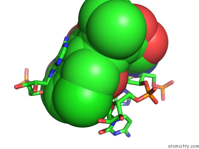
Mono view
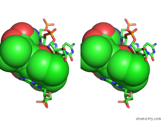
Stereo pair view
Mono view
Stereo pair view
A full contact list of Chlorine with other atoms in the Cl binding
site number 1 of Crystal Structure of An in Vitro Evolved Tetracycline Aptamer and Artificial Riboswitch within 5.0Å range:
|
Reference:
H.Xiao,
T.E.Edwards,
A.R.Ferre-D'amare.
Structural Basis For Specific, High-Affinity Tetracycline Binding By An in Vitro Evolved Aptamer and Artificial Riboswitch Chem.Biol. V. 15 1125 2008.
ISSN: ISSN 1074-5521
PubMed: 18940672
DOI: 10.1016/J.CHEMBIOL.2008.09.004
Page generated: Fri Jul 11 04:48:29 2025
ISSN: ISSN 1074-5521
PubMed: 18940672
DOI: 10.1016/J.CHEMBIOL.2008.09.004
Last articles
Cl in 8AV4Cl in 8AV3
Cl in 8AU8
Cl in 8AUY
Cl in 8AUS
Cl in 8AU2
Cl in 8AU5
Cl in 8ATY
Cl in 8ATZ
Cl in 8ATS