Chlorine »
PDB 3ecg-3es1 »
3ejd »
Chlorine in PDB 3ejd: Crystal Structure of P450BIOI in Complex with Hexadec-9Z-Enoic Acid Ligated Acyl Carrier Protein
Protein crystallography data
The structure of Crystal Structure of P450BIOI in Complex with Hexadec-9Z-Enoic Acid Ligated Acyl Carrier Protein, PDB code: 3ejd
was solved by
M.J.Cryle,
I.Schlichting,
with X-Ray Crystallography technique. A brief refinement statistics is given in the table below:
| Resolution Low / High (Å) | 20.00 / 2.10 |
| Space group | P 1 |
| Cell size a, b, c (Å), α, β, γ (°) | 61.300, 92.100, 107.700, 109.00, 89.20, 90.10 |
| R / Rfree (%) | 24.1 / 28.5 |
Other elements in 3ejd:
The structure of Crystal Structure of P450BIOI in Complex with Hexadec-9Z-Enoic Acid Ligated Acyl Carrier Protein also contains other interesting chemical elements:
| Iron | (Fe) | 4 atoms |
Chlorine Binding Sites:
The binding sites of Chlorine atom in the Crystal Structure of P450BIOI in Complex with Hexadec-9Z-Enoic Acid Ligated Acyl Carrier Protein
(pdb code 3ejd). This binding sites where shown within
5.0 Angstroms radius around Chlorine atom.
In total 2 binding sites of Chlorine where determined in the Crystal Structure of P450BIOI in Complex with Hexadec-9Z-Enoic Acid Ligated Acyl Carrier Protein, PDB code: 3ejd:
Jump to Chlorine binding site number: 1; 2;
In total 2 binding sites of Chlorine where determined in the Crystal Structure of P450BIOI in Complex with Hexadec-9Z-Enoic Acid Ligated Acyl Carrier Protein, PDB code: 3ejd:
Jump to Chlorine binding site number: 1; 2;
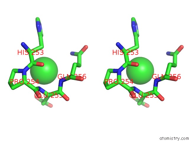
Chlorine binding site 1 out of 2 in 3ejd
Go back to
Chlorine binding site 1 out
of 2 in the Crystal Structure of P450BIOI in Complex with Hexadec-9Z-Enoic Acid Ligated Acyl Carrier Protein

Mono view

Stereo pair view
Mono view
Stereo pair view
A full contact list of Chlorine with other atoms in the Cl binding
site number 1 of Crystal Structure of P450BIOI in Complex with Hexadec-9Z-Enoic Acid Ligated Acyl Carrier Protein within 5.0Å range:
|
Chlorine binding site 2 out of 2 in 3ejd
Go back to
Chlorine binding site 2 out
of 2 in the Crystal Structure of P450BIOI in Complex with Hexadec-9Z-Enoic Acid Ligated Acyl Carrier Protein

Mono view
Stereo pair view
Mono view
Stereo pair view
A full contact list of Chlorine with other atoms in the Cl binding
site number 2 of Crystal Structure of P450BIOI in Complex with Hexadec-9Z-Enoic Acid Ligated Acyl Carrier Protein within 5.0Å range:
|
Reference:
M.J.Cryle,
I.Schlichting.
Structural Insights From A P450 Carrier Protein Complex Reveal How Specificity Is Achieved in the P450(Bioi) Acp Complex. Proc.Natl.Acad.Sci.Usa V. 105 15696 2008.
ISSN: ISSN 0027-8424
PubMed: 18838690
DOI: 10.1073/PNAS.0805983105
Page generated: Sat Jul 20 18:51:41 2024
ISSN: ISSN 0027-8424
PubMed: 18838690
DOI: 10.1073/PNAS.0805983105
Last articles
Zn in 9J0NZn in 9J0O
Zn in 9J0P
Zn in 9FJX
Zn in 9EKB
Zn in 9C0F
Zn in 9CAH
Zn in 9CH0
Zn in 9CH3
Zn in 9CH1