Chlorine »
PDB 3es4-3f68 »
3ezw »
Chlorine in PDB 3ezw: Crystal Structure of A Hyperactive Escherichia Coli Glycerol Kinase Mutant GLY230 --> Asp Obtained Using Microfluidic Crystallization Devices
Enzymatic activity of Crystal Structure of A Hyperactive Escherichia Coli Glycerol Kinase Mutant GLY230 --> Asp Obtained Using Microfluidic Crystallization Devices
All present enzymatic activity of Crystal Structure of A Hyperactive Escherichia Coli Glycerol Kinase Mutant GLY230 --> Asp Obtained Using Microfluidic Crystallization Devices:
2.7.1.30;
2.7.1.30;
Protein crystallography data
The structure of Crystal Structure of A Hyperactive Escherichia Coli Glycerol Kinase Mutant GLY230 --> Asp Obtained Using Microfluidic Crystallization Devices, PDB code: 3ezw
was solved by
M.J.Anderson,
B.Delabarre,
P.Dunten,
A.T.Brunger,
S.R.Quake,
with X-Ray Crystallography technique. A brief refinement statistics is given in the table below:
| Resolution Low / High (Å) | 20.00 / 2.00 |
| Space group | P 1 21 1 |
| Cell size a, b, c (Å), α, β, γ (°) | 91.116, 114.260, 212.624, 90.00, 91.15, 90.00 |
| R / Rfree (%) | 16.7 / 22.5 |
Other elements in 3ezw:
The structure of Crystal Structure of A Hyperactive Escherichia Coli Glycerol Kinase Mutant GLY230 --> Asp Obtained Using Microfluidic Crystallization Devices also contains other interesting chemical elements:
| Magnesium | (Mg) | 1 atom |
Chlorine Binding Sites:
The binding sites of Chlorine atom in the Crystal Structure of A Hyperactive Escherichia Coli Glycerol Kinase Mutant GLY230 --> Asp Obtained Using Microfluidic Crystallization Devices
(pdb code 3ezw). This binding sites where shown within
5.0 Angstroms radius around Chlorine atom.
In total 8 binding sites of Chlorine where determined in the Crystal Structure of A Hyperactive Escherichia Coli Glycerol Kinase Mutant GLY230 --> Asp Obtained Using Microfluidic Crystallization Devices, PDB code: 3ezw:
Jump to Chlorine binding site number: 1; 2; 3; 4; 5; 6; 7; 8;
In total 8 binding sites of Chlorine where determined in the Crystal Structure of A Hyperactive Escherichia Coli Glycerol Kinase Mutant GLY230 --> Asp Obtained Using Microfluidic Crystallization Devices, PDB code: 3ezw:
Jump to Chlorine binding site number: 1; 2; 3; 4; 5; 6; 7; 8;
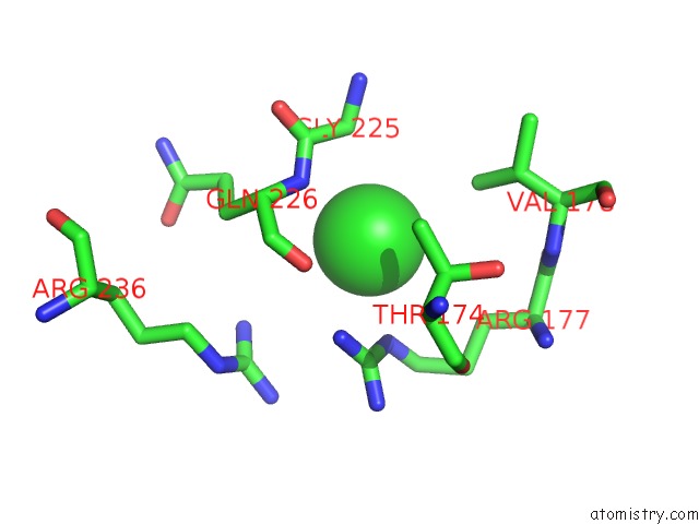
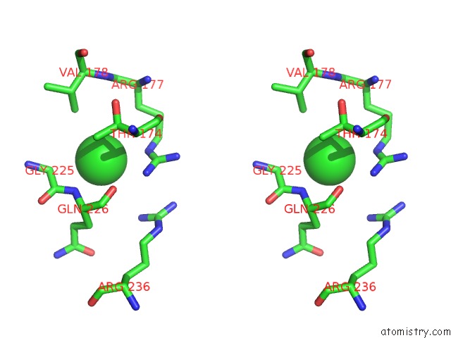
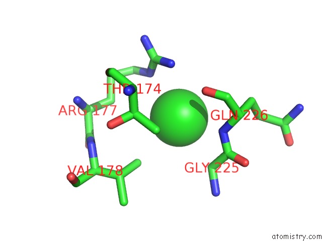
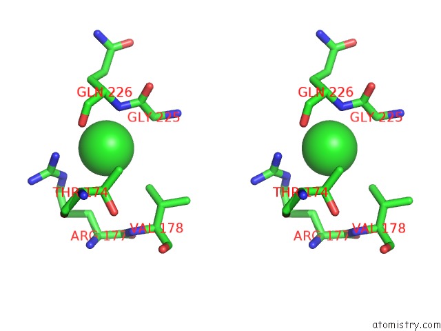
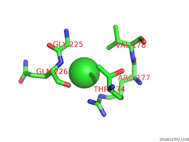
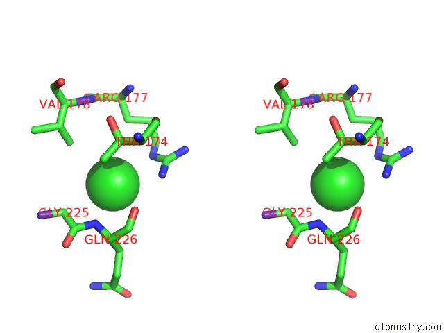
Chlorine binding site 1 out of 8 in 3ezw
Go back to
Chlorine binding site 1 out
of 8 in the Crystal Structure of A Hyperactive Escherichia Coli Glycerol Kinase Mutant GLY230 --> Asp Obtained Using Microfluidic Crystallization Devices
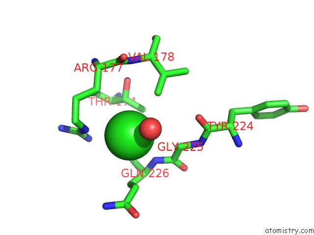
Mono view
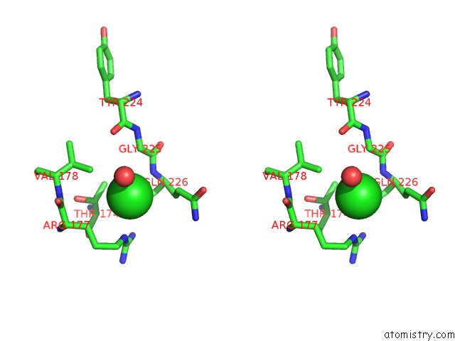
Stereo pair view
Mono view
Stereo pair view
A full contact list of Chlorine with other atoms in the Cl binding
site number 1 of Crystal Structure of A Hyperactive Escherichia Coli Glycerol Kinase Mutant GLY230 --> Asp Obtained Using Microfluidic Crystallization Devices within 5.0Å range:
|
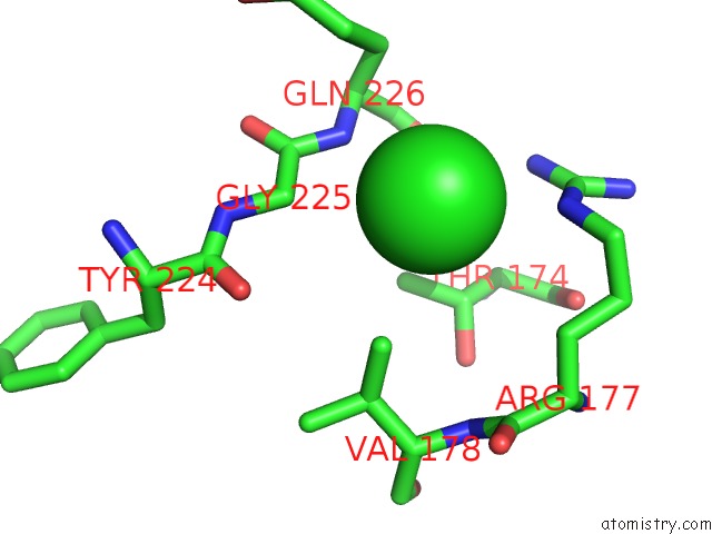
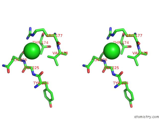
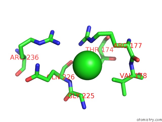
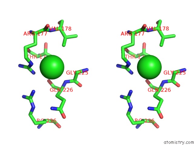
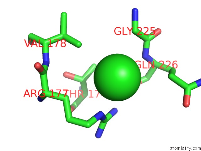
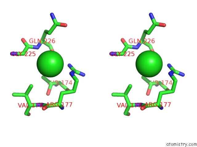
Chlorine binding site 2 out of 8 in 3ezw
Go back to
Chlorine binding site 2 out
of 8 in the Crystal Structure of A Hyperactive Escherichia Coli Glycerol Kinase Mutant GLY230 --> Asp Obtained Using Microfluidic Crystallization Devices
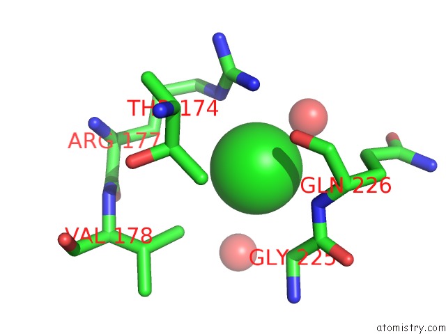
Mono view
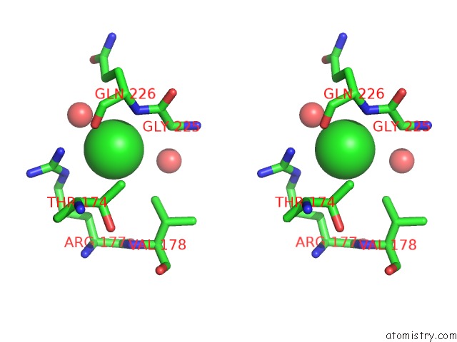
Stereo pair view
Mono view
Stereo pair view
A full contact list of Chlorine with other atoms in the Cl binding
site number 2 of Crystal Structure of A Hyperactive Escherichia Coli Glycerol Kinase Mutant GLY230 --> Asp Obtained Using Microfluidic Crystallization Devices within 5.0Å range:
|
Chlorine binding site 3 out of 8 in 3ezw
Go back to
Chlorine binding site 3 out
of 8 in the Crystal Structure of A Hyperactive Escherichia Coli Glycerol Kinase Mutant GLY230 --> Asp Obtained Using Microfluidic Crystallization Devices
Mono view
Stereo pair view
Mono view
Stereo pair view
A full contact list of Chlorine with other atoms in the Cl binding
site number 3 of Crystal Structure of A Hyperactive Escherichia Coli Glycerol Kinase Mutant GLY230 --> Asp Obtained Using Microfluidic Crystallization Devices within 5.0Å range:
|
Chlorine binding site 4 out of 8 in 3ezw
Go back to
Chlorine binding site 4 out
of 8 in the Crystal Structure of A Hyperactive Escherichia Coli Glycerol Kinase Mutant GLY230 --> Asp Obtained Using Microfluidic Crystallization Devices
Mono view
Stereo pair view
Mono view
Stereo pair view
A full contact list of Chlorine with other atoms in the Cl binding
site number 4 of Crystal Structure of A Hyperactive Escherichia Coli Glycerol Kinase Mutant GLY230 --> Asp Obtained Using Microfluidic Crystallization Devices within 5.0Å range:
|
Chlorine binding site 5 out of 8 in 3ezw
Go back to
Chlorine binding site 5 out
of 8 in the Crystal Structure of A Hyperactive Escherichia Coli Glycerol Kinase Mutant GLY230 --> Asp Obtained Using Microfluidic Crystallization Devices
Mono view
Stereo pair view
Mono view
Stereo pair view
A full contact list of Chlorine with other atoms in the Cl binding
site number 5 of Crystal Structure of A Hyperactive Escherichia Coli Glycerol Kinase Mutant GLY230 --> Asp Obtained Using Microfluidic Crystallization Devices within 5.0Å range:
|
Chlorine binding site 6 out of 8 in 3ezw
Go back to
Chlorine binding site 6 out
of 8 in the Crystal Structure of A Hyperactive Escherichia Coli Glycerol Kinase Mutant GLY230 --> Asp Obtained Using Microfluidic Crystallization Devices
Mono view
Stereo pair view
Mono view
Stereo pair view
A full contact list of Chlorine with other atoms in the Cl binding
site number 6 of Crystal Structure of A Hyperactive Escherichia Coli Glycerol Kinase Mutant GLY230 --> Asp Obtained Using Microfluidic Crystallization Devices within 5.0Å range:
|
Chlorine binding site 7 out of 8 in 3ezw
Go back to
Chlorine binding site 7 out
of 8 in the Crystal Structure of A Hyperactive Escherichia Coli Glycerol Kinase Mutant GLY230 --> Asp Obtained Using Microfluidic Crystallization Devices
Mono view
Stereo pair view
Mono view
Stereo pair view
A full contact list of Chlorine with other atoms in the Cl binding
site number 7 of Crystal Structure of A Hyperactive Escherichia Coli Glycerol Kinase Mutant GLY230 --> Asp Obtained Using Microfluidic Crystallization Devices within 5.0Å range:
|
Chlorine binding site 8 out of 8 in 3ezw
Go back to
Chlorine binding site 8 out
of 8 in the Crystal Structure of A Hyperactive Escherichia Coli Glycerol Kinase Mutant GLY230 --> Asp Obtained Using Microfluidic Crystallization Devices
Mono view
Stereo pair view
Mono view
Stereo pair view
A full contact list of Chlorine with other atoms in the Cl binding
site number 8 of Crystal Structure of A Hyperactive Escherichia Coli Glycerol Kinase Mutant GLY230 --> Asp Obtained Using Microfluidic Crystallization Devices within 5.0Å range:
|
Reference:
M.J.Anderson,
B.Delabarre,
A.Raghunathan,
B.O.Palsson,
A.T.Brunger,
S.R.Quake.
Crystal Structure of A Hyperactive Escherichia Coli Glycerol Kinase Mutant GLY230 --> Asp Obtained Using Microfluidic Crystallization Devices. Biochemistry V. 46 5722 2007.
ISSN: ISSN 0006-2960
PubMed: 17441732
DOI: 10.1021/BI700096P
Page generated: Sat Jul 20 19:03:08 2024
ISSN: ISSN 0006-2960
PubMed: 17441732
DOI: 10.1021/BI700096P
Last articles
Zn in 9J0NZn in 9J0O
Zn in 9J0P
Zn in 9FJX
Zn in 9EKB
Zn in 9C0F
Zn in 9CAH
Zn in 9CH0
Zn in 9CH3
Zn in 9CH1