Chlorine »
PDB 3f75-3fee »
3fea »
Chlorine in PDB 3fea: Crystal Structure of Hdmx Bound to the P53-Peptidomimetic Ac-Phe-Met- Aib-Pmp-6-Cl-Trp-Glu-AC3C-Leu-NH2 at 1.33A
Protein crystallography data
The structure of Crystal Structure of Hdmx Bound to the P53-Peptidomimetic Ac-Phe-Met- Aib-Pmp-6-Cl-Trp-Glu-AC3C-Leu-NH2 at 1.33A, PDB code: 3fea
was solved by
J.Kallen,
with X-Ray Crystallography technique. A brief refinement statistics is given in the table below:
| Resolution Low / High (Å) | 20.00 / 1.33 |
| Space group | P 41 |
| Cell size a, b, c (Å), α, β, γ (°) | 42.249, 42.249, 70.449, 90.00, 90.00, 90.00 |
| R / Rfree (%) | 19.3 / 20.6 |
Chlorine Binding Sites:
The binding sites of Chlorine atom in the Crystal Structure of Hdmx Bound to the P53-Peptidomimetic Ac-Phe-Met- Aib-Pmp-6-Cl-Trp-Glu-AC3C-Leu-NH2 at 1.33A
(pdb code 3fea). This binding sites where shown within
5.0 Angstroms radius around Chlorine atom.
In total 2 binding sites of Chlorine where determined in the Crystal Structure of Hdmx Bound to the P53-Peptidomimetic Ac-Phe-Met- Aib-Pmp-6-Cl-Trp-Glu-AC3C-Leu-NH2 at 1.33A, PDB code: 3fea:
Jump to Chlorine binding site number: 1; 2;
In total 2 binding sites of Chlorine where determined in the Crystal Structure of Hdmx Bound to the P53-Peptidomimetic Ac-Phe-Met- Aib-Pmp-6-Cl-Trp-Glu-AC3C-Leu-NH2 at 1.33A, PDB code: 3fea:
Jump to Chlorine binding site number: 1; 2;
Chlorine binding site 1 out of 2 in 3fea
Go back to
Chlorine binding site 1 out
of 2 in the Crystal Structure of Hdmx Bound to the P53-Peptidomimetic Ac-Phe-Met- Aib-Pmp-6-Cl-Trp-Glu-AC3C-Leu-NH2 at 1.33A
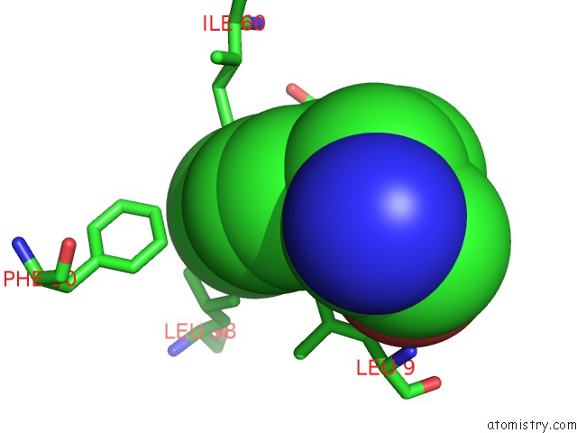
Mono view
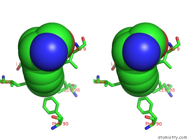
Stereo pair view
Mono view
Stereo pair view
A full contact list of Chlorine with other atoms in the Cl binding
site number 1 of Crystal Structure of Hdmx Bound to the P53-Peptidomimetic Ac-Phe-Met- Aib-Pmp-6-Cl-Trp-Glu-AC3C-Leu-NH2 at 1.33A within 5.0Å range:
|
Chlorine binding site 2 out of 2 in 3fea
Go back to
Chlorine binding site 2 out
of 2 in the Crystal Structure of Hdmx Bound to the P53-Peptidomimetic Ac-Phe-Met- Aib-Pmp-6-Cl-Trp-Glu-AC3C-Leu-NH2 at 1.33A
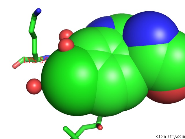
Mono view
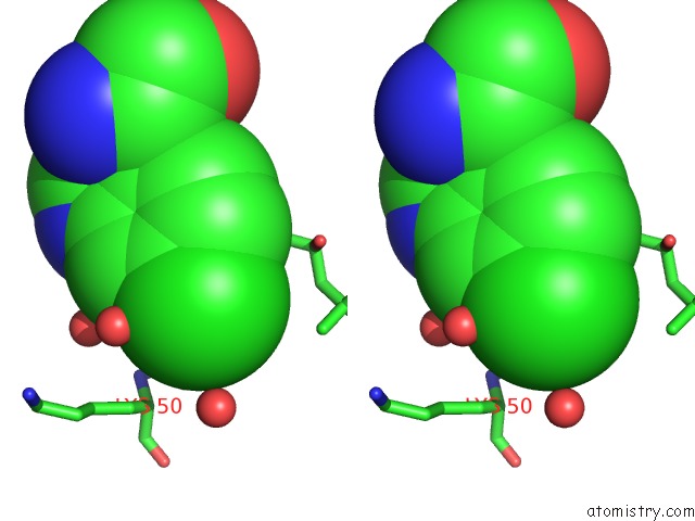
Stereo pair view
Mono view
Stereo pair view
A full contact list of Chlorine with other atoms in the Cl binding
site number 2 of Crystal Structure of Hdmx Bound to the P53-Peptidomimetic Ac-Phe-Met- Aib-Pmp-6-Cl-Trp-Glu-AC3C-Leu-NH2 at 1.33A within 5.0Å range:
|
Reference:
J.Kallen,
A.Goepfert,
A.Blechschmidt,
A.Izaac,
M.Geiser,
G.Tavares,
P.Ramage,
P.Furet,
K.Masuya,
J.Lisztwan.
Crystal Structures of Human Mdmx (Hdmx) in Complex with P53 Peptide Analogues Reveal Surprising Conformational Changes J.Biol.Chem. V. 284 8812 2009.
ISSN: ISSN 0021-9258
PubMed: 19153082
DOI: 10.1074/JBC.M809096200
Page generated: Sat Jul 20 19:20:29 2024
ISSN: ISSN 0021-9258
PubMed: 19153082
DOI: 10.1074/JBC.M809096200
Last articles
Zn in 9J0NZn in 9J0O
Zn in 9J0P
Zn in 9FJX
Zn in 9EKB
Zn in 9C0F
Zn in 9CAH
Zn in 9CH0
Zn in 9CH3
Zn in 9CH1