Chlorine »
PDB 3fei-3fpl »
3fen »
Chlorine in PDB 3fen: Crystal Structure of the R132K:R111L:A32E Mutant of Cellular Retinoic Acid-Binding Protein II at 1.56 Angstrom Resolution
Protein crystallography data
The structure of Crystal Structure of the R132K:R111L:A32E Mutant of Cellular Retinoic Acid-Binding Protein II at 1.56 Angstrom Resolution, PDB code: 3fen
was solved by
X.Jia,
J.H.Geiger,
with X-Ray Crystallography technique. A brief refinement statistics is given in the table below:
| Resolution Low / High (Å) | 25.91 / 1.56 |
| Space group | P 1 |
| Cell size a, b, c (Å), α, β, γ (°) | 34.692, 37.071, 58.549, 102.55, 106.37, 92.87 |
| R / Rfree (%) | 15.4 / 19.8 |
Chlorine Binding Sites:
The binding sites of Chlorine atom in the Crystal Structure of the R132K:R111L:A32E Mutant of Cellular Retinoic Acid-Binding Protein II at 1.56 Angstrom Resolution
(pdb code 3fen). This binding sites where shown within
5.0 Angstroms radius around Chlorine atom.
In total 3 binding sites of Chlorine where determined in the Crystal Structure of the R132K:R111L:A32E Mutant of Cellular Retinoic Acid-Binding Protein II at 1.56 Angstrom Resolution, PDB code: 3fen:
Jump to Chlorine binding site number: 1; 2; 3;
In total 3 binding sites of Chlorine where determined in the Crystal Structure of the R132K:R111L:A32E Mutant of Cellular Retinoic Acid-Binding Protein II at 1.56 Angstrom Resolution, PDB code: 3fen:
Jump to Chlorine binding site number: 1; 2; 3;
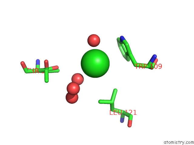
Chlorine binding site 1 out of 3 in 3fen
Go back to
Chlorine binding site 1 out
of 3 in the Crystal Structure of the R132K:R111L:A32E Mutant of Cellular Retinoic Acid-Binding Protein II at 1.56 Angstrom Resolution
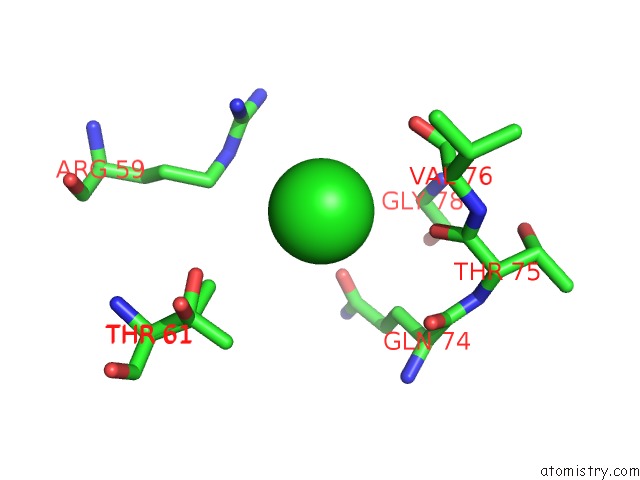
Mono view
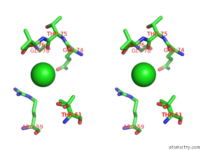
Stereo pair view
Mono view
Stereo pair view
A full contact list of Chlorine with other atoms in the Cl binding
site number 1 of Crystal Structure of the R132K:R111L:A32E Mutant of Cellular Retinoic Acid-Binding Protein II at 1.56 Angstrom Resolution within 5.0Å range:
|
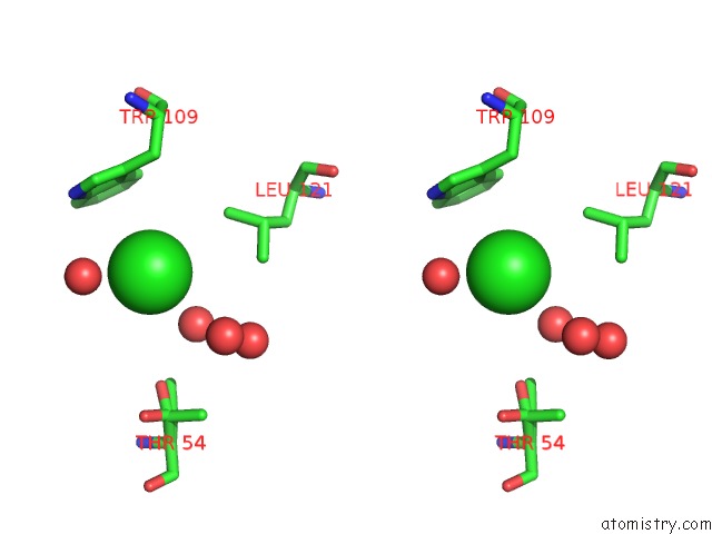
Chlorine binding site 2 out of 3 in 3fen
Go back to
Chlorine binding site 2 out
of 3 in the Crystal Structure of the R132K:R111L:A32E Mutant of Cellular Retinoic Acid-Binding Protein II at 1.56 Angstrom Resolution
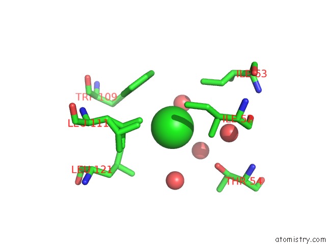
Mono view
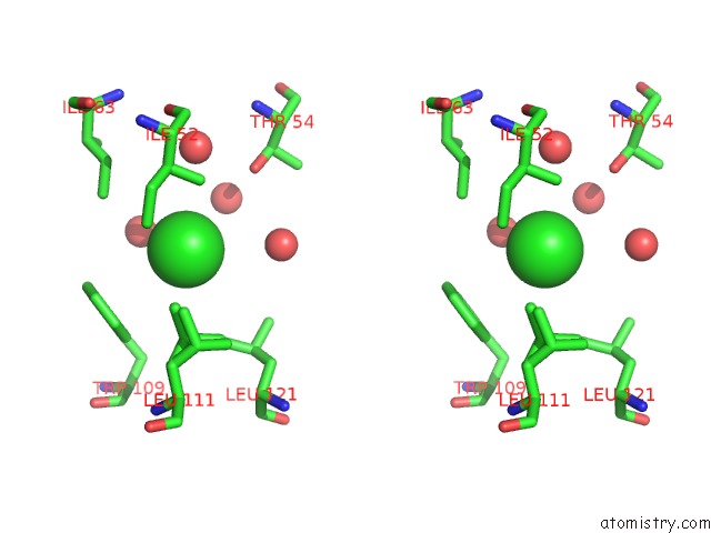
Stereo pair view
Mono view
Stereo pair view
A full contact list of Chlorine with other atoms in the Cl binding
site number 2 of Crystal Structure of the R132K:R111L:A32E Mutant of Cellular Retinoic Acid-Binding Protein II at 1.56 Angstrom Resolution within 5.0Å range:
|
Chlorine binding site 3 out of 3 in 3fen
Go back to
Chlorine binding site 3 out
of 3 in the Crystal Structure of the R132K:R111L:A32E Mutant of Cellular Retinoic Acid-Binding Protein II at 1.56 Angstrom Resolution
Mono view
Stereo pair view
Mono view
Stereo pair view
A full contact list of Chlorine with other atoms in the Cl binding
site number 3 of Crystal Structure of the R132K:R111L:A32E Mutant of Cellular Retinoic Acid-Binding Protein II at 1.56 Angstrom Resolution within 5.0Å range:
|
Reference:
X.Jia,
K.S.Lee,
C.Vasileiou,
B.Borhan,
J.H.Geiger.
Crystal Structures of Apo Cellular Retinoic Acid-Binding Protein II Mutants: Structural Integrity Investigated Through Multiple Site Mutations To Be Published.
Page generated: Sat Jul 20 19:21:51 2024
Last articles
Zn in 9J0NZn in 9J0O
Zn in 9J0P
Zn in 9FJX
Zn in 9EKB
Zn in 9C0F
Zn in 9CAH
Zn in 9CH0
Zn in 9CH3
Zn in 9CH1