Chlorine »
PDB 3fei-3fpl »
3fo5 »
Chlorine in PDB 3fo5: Human Start Domain of Acyl-Coenzyme A Thioesterase 11 (ACOT11)
Protein crystallography data
The structure of Human Start Domain of Acyl-Coenzyme A Thioesterase 11 (ACOT11), PDB code: 3fo5
was solved by
M.I.Siponen,
L.Lehtio,
C.H.Arrowsmith,
H.Berglund,
C.Bountra,
R.Collins,
L.G.Dahlgren,
A.M.Edwards,
S.Flodin,
A.Flores,
S.Graslund,
M.Hammarstrom,
A.Johansson,
I.Johansson,
T.Karlberg,
T.Kotenyova,
M.Moche,
M.E.Nilsson,
P.Nordlund,
T.Nyman,
C.Persson,
J.Sagemark,
A.G.Thorsell,
L.Tresaugues,
S.Van-Den-Berg,
J.Weigelt,
M.Welin,
M.Wikstrom,
M.Wisniewska,
H.Shueler,
Structural Genomics Consortium (Sgc),
with X-Ray Crystallography technique. A brief refinement statistics is given in the table below:
| Resolution Low / High (Å) | 36.47 / 2.00 |
| Space group | C 2 2 21 |
| Cell size a, b, c (Å), α, β, γ (°) | 52.440, 130.080, 165.230, 90.00, 90.00, 90.00 |
| R / Rfree (%) | 20.2 / 25.1 |
Chlorine Binding Sites:
The binding sites of Chlorine atom in the Human Start Domain of Acyl-Coenzyme A Thioesterase 11 (ACOT11)
(pdb code 3fo5). This binding sites where shown within
5.0 Angstroms radius around Chlorine atom.
In total only one binding site of Chlorine was determined in the Human Start Domain of Acyl-Coenzyme A Thioesterase 11 (ACOT11), PDB code: 3fo5:
In total only one binding site of Chlorine was determined in the Human Start Domain of Acyl-Coenzyme A Thioesterase 11 (ACOT11), PDB code: 3fo5:
Chlorine binding site 1 out of 1 in 3fo5
Go back to
Chlorine binding site 1 out
of 1 in the Human Start Domain of Acyl-Coenzyme A Thioesterase 11 (ACOT11)
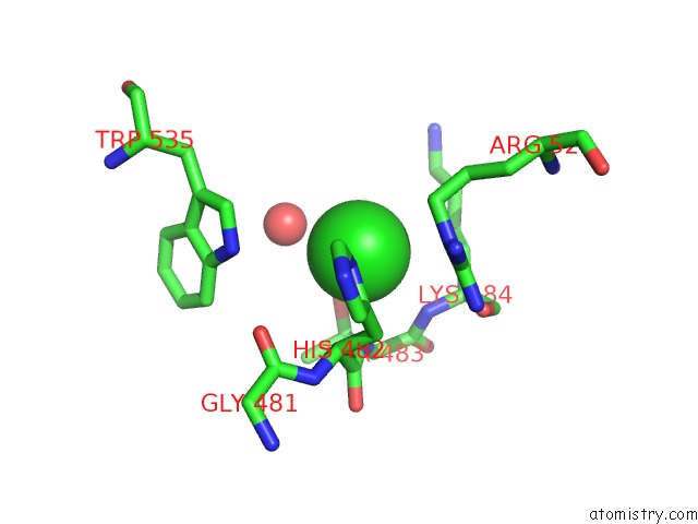
Mono view
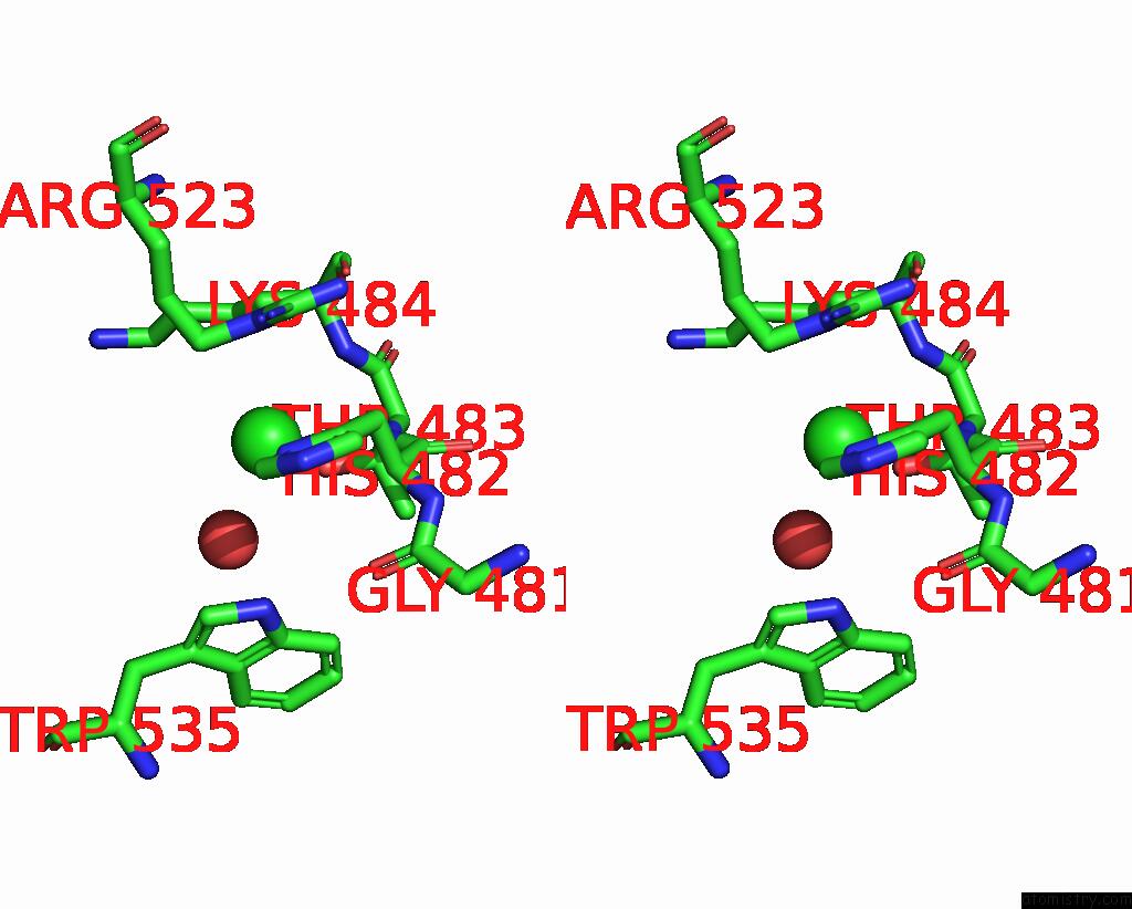
Stereo pair view
Mono view
Stereo pair view
A full contact list of Chlorine with other atoms in the Cl binding
site number 1 of Human Start Domain of Acyl-Coenzyme A Thioesterase 11 (ACOT11) within 5.0Å range:
|
Reference:
A.G.Thorsell,
W.H.Lee,
C.Persson,
M.I.Siponen,
M.Nilsson,
R.D.Busam,
T.Kotenyova,
H.Schuler,
L.Lehtio.
Comparative Structural Analysis of Lipid Binding Start Domains. Plos One V. 6 19521 2011.
ISSN: ESSN 1932-6203
PubMed: 21738568
DOI: 10.1371/JOURNAL.PONE.0019521
Page generated: Sat Jul 20 19:31:25 2024
ISSN: ESSN 1932-6203
PubMed: 21738568
DOI: 10.1371/JOURNAL.PONE.0019521
Last articles
Zn in 9J0NZn in 9J0O
Zn in 9J0P
Zn in 9FJX
Zn in 9EKB
Zn in 9C0F
Zn in 9CAH
Zn in 9CH0
Zn in 9CH3
Zn in 9CH1