Chlorine »
PDB 3fzy-3g72 »
3g1v »
Chlorine in PDB 3g1v: Crystal Structure of the Mutant D70G of Orotidine 5'- Monophosphate Decarboxylase From Methanobacterium Thermoautotrophicum Complexed with 5-Fluorouridine 5'- Monophosphate
Enzymatic activity of Crystal Structure of the Mutant D70G of Orotidine 5'- Monophosphate Decarboxylase From Methanobacterium Thermoautotrophicum Complexed with 5-Fluorouridine 5'- Monophosphate
All present enzymatic activity of Crystal Structure of the Mutant D70G of Orotidine 5'- Monophosphate Decarboxylase From Methanobacterium Thermoautotrophicum Complexed with 5-Fluorouridine 5'- Monophosphate:
4.1.1.23;
4.1.1.23;
Protein crystallography data
The structure of Crystal Structure of the Mutant D70G of Orotidine 5'- Monophosphate Decarboxylase From Methanobacterium Thermoautotrophicum Complexed with 5-Fluorouridine 5'- Monophosphate, PDB code: 3g1v
was solved by
A.A.Fedorov,
E.V.Fedorov,
K.K.Chan,
J.A.Gerlt,
S.C.Almo,
with X-Ray Crystallography technique. A brief refinement statistics is given in the table below:
| Resolution Low / High (Å) | 24.93 / 1.30 |
| Space group | P 41 |
| Cell size a, b, c (Å), α, β, γ (°) | 56.620, 56.620, 127.443, 90.00, 90.00, 90.00 |
| R / Rfree (%) | 19.3 / 21.1 |
Other elements in 3g1v:
The structure of Crystal Structure of the Mutant D70G of Orotidine 5'- Monophosphate Decarboxylase From Methanobacterium Thermoautotrophicum Complexed with 5-Fluorouridine 5'- Monophosphate also contains other interesting chemical elements:
| Fluorine | (F) | 2 atoms |
Chlorine Binding Sites:
The binding sites of Chlorine atom in the Crystal Structure of the Mutant D70G of Orotidine 5'- Monophosphate Decarboxylase From Methanobacterium Thermoautotrophicum Complexed with 5-Fluorouridine 5'- Monophosphate
(pdb code 3g1v). This binding sites where shown within
5.0 Angstroms radius around Chlorine atom.
In total 2 binding sites of Chlorine where determined in the Crystal Structure of the Mutant D70G of Orotidine 5'- Monophosphate Decarboxylase From Methanobacterium Thermoautotrophicum Complexed with 5-Fluorouridine 5'- Monophosphate, PDB code: 3g1v:
Jump to Chlorine binding site number: 1; 2;
In total 2 binding sites of Chlorine where determined in the Crystal Structure of the Mutant D70G of Orotidine 5'- Monophosphate Decarboxylase From Methanobacterium Thermoautotrophicum Complexed with 5-Fluorouridine 5'- Monophosphate, PDB code: 3g1v:
Jump to Chlorine binding site number: 1; 2;
Chlorine binding site 1 out of 2 in 3g1v
Go back to
Chlorine binding site 1 out
of 2 in the Crystal Structure of the Mutant D70G of Orotidine 5'- Monophosphate Decarboxylase From Methanobacterium Thermoautotrophicum Complexed with 5-Fluorouridine 5'- Monophosphate
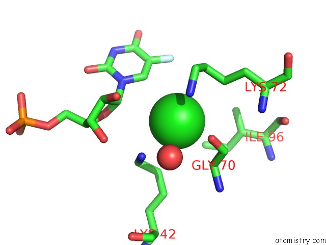
Mono view
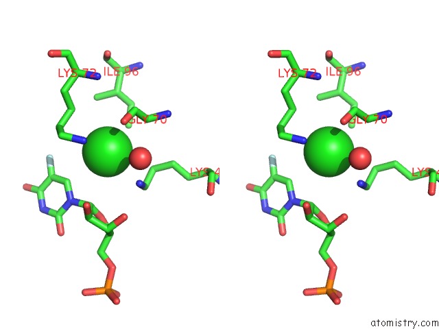
Stereo pair view
Mono view
Stereo pair view
A full contact list of Chlorine with other atoms in the Cl binding
site number 1 of Crystal Structure of the Mutant D70G of Orotidine 5'- Monophosphate Decarboxylase From Methanobacterium Thermoautotrophicum Complexed with 5-Fluorouridine 5'- Monophosphate within 5.0Å range:
|
Chlorine binding site 2 out of 2 in 3g1v
Go back to
Chlorine binding site 2 out
of 2 in the Crystal Structure of the Mutant D70G of Orotidine 5'- Monophosphate Decarboxylase From Methanobacterium Thermoautotrophicum Complexed with 5-Fluorouridine 5'- Monophosphate
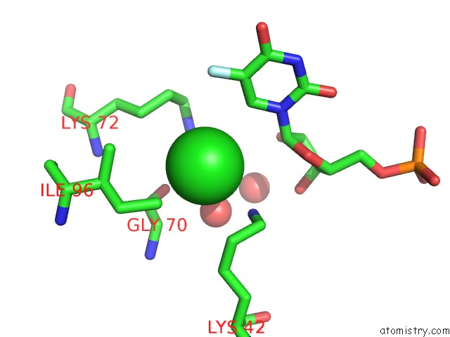
Mono view
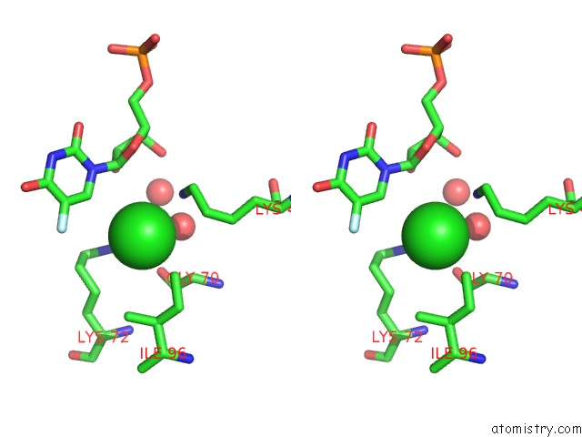
Stereo pair view
Mono view
Stereo pair view
A full contact list of Chlorine with other atoms in the Cl binding
site number 2 of Crystal Structure of the Mutant D70G of Orotidine 5'- Monophosphate Decarboxylase From Methanobacterium Thermoautotrophicum Complexed with 5-Fluorouridine 5'- Monophosphate within 5.0Å range:
|
Reference:
K.K.Chan,
B.M.Wood,
A.A.Fedorov,
E.V.Fedorov,
H.J.Imker,
T.L.Amyes,
J.P.Richard,
S.C.Almo,
J.A.Gerlt.
Mechanism of the Orotidine 5'-Monophosphate Decarboxylase-Catalyzed Reaction: Evidence For Substrate Destabilization. Biochemistry V. 48 5518 2009.
ISSN: ISSN 0006-2960
PubMed: 19435314
DOI: 10.1021/BI900623R
Page generated: Fri Jul 11 05:25:23 2025
ISSN: ISSN 0006-2960
PubMed: 19435314
DOI: 10.1021/BI900623R
Last articles
Fe in 2YXOFe in 2YRS
Fe in 2YXC
Fe in 2YNM
Fe in 2YVJ
Fe in 2YP1
Fe in 2YU2
Fe in 2YU1
Fe in 2YQB
Fe in 2YOO