Chlorine »
PDB 3gky-3gxg »
3gog »
Chlorine in PDB 3gog: Guanine Riboswitch A21G,U75C Mutant Bound to 6-Chloroguanine
Protein crystallography data
The structure of Guanine Riboswitch A21G,U75C Mutant Bound to 6-Chloroguanine, PDB code: 3gog
was solved by
S.D.Gilbert,
R.T.Batey,
with X-Ray Crystallography technique. A brief refinement statistics is given in the table below:
| Resolution Low / High (Å) | 19.97 / 2.10 |
| Space group | C 1 2 1 |
| Cell size a, b, c (Å), α, β, γ (°) | 132.510, 35.120, 41.760, 90.00, 90.57, 90.00 |
| R / Rfree (%) | 22 / 25.6 |
Other elements in 3gog:
The structure of Guanine Riboswitch A21G,U75C Mutant Bound to 6-Chloroguanine also contains other interesting chemical elements:
| Cobalt | (Co) | 8 atoms |
Chlorine Binding Sites:
The binding sites of Chlorine atom in the Guanine Riboswitch A21G,U75C Mutant Bound to 6-Chloroguanine
(pdb code 3gog). This binding sites where shown within
5.0 Angstroms radius around Chlorine atom.
In total only one binding site of Chlorine was determined in the Guanine Riboswitch A21G,U75C Mutant Bound to 6-Chloroguanine, PDB code: 3gog:
In total only one binding site of Chlorine was determined in the Guanine Riboswitch A21G,U75C Mutant Bound to 6-Chloroguanine, PDB code: 3gog:
Chlorine binding site 1 out of 1 in 3gog
Go back to
Chlorine binding site 1 out
of 1 in the Guanine Riboswitch A21G,U75C Mutant Bound to 6-Chloroguanine
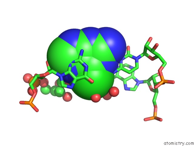
Mono view
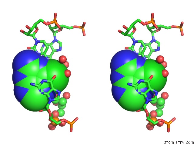
Stereo pair view
Mono view
Stereo pair view
A full contact list of Chlorine with other atoms in the Cl binding
site number 1 of Guanine Riboswitch A21G,U75C Mutant Bound to 6-Chloroguanine within 5.0Å range:
|
Reference:
S.D.Gilbert,
F.E.Reyes,
A.L.Edwards,
R.T.Batey.
Adaptive Ligand Binding By the Purine Riboswitch in the Recognition of Guanine and Adenine Analogs Structure V. 17 857 2009.
ISSN: ISSN 0969-2126
PubMed: 19523903
DOI: 10.1016/J.STR.2009.04.009
Page generated: Fri Jul 11 05:44:14 2025
ISSN: ISSN 0969-2126
PubMed: 19523903
DOI: 10.1016/J.STR.2009.04.009
Last articles
F in 4JVSF in 4JVR
F in 4JU1
F in 4JUO
F in 4JVE
F in 4JU4
F in 4JTZ
F in 4JTY
F in 4JTW
F in 4JSX