Chlorine »
PDB 3hd1-3hl7 »
3hfs »
Chlorine in PDB 3hfs: Structure of Apo Anthocyanidin Reductase From Vitis Vinifera
Enzymatic activity of Structure of Apo Anthocyanidin Reductase From Vitis Vinifera
All present enzymatic activity of Structure of Apo Anthocyanidin Reductase From Vitis Vinifera:
1.3.1.77;
1.3.1.77;
Protein crystallography data
The structure of Structure of Apo Anthocyanidin Reductase From Vitis Vinifera, PDB code: 3hfs
was solved by
M.Gargouri,
C.Mauge,
B.Langlois D'estaintot,
T.Granier,
C.Manigan,
B.Gallois,
with X-Ray Crystallography technique. A brief refinement statistics is given in the table below:
| Resolution Low / High (Å) | 79.31 / 3.17 |
| Space group | P 1 21 1 |
| Cell size a, b, c (Å), α, β, γ (°) | 84.518, 51.012, 86.113, 90.00, 110.31, 90.00 |
| R / Rfree (%) | 22.1 / 29.9 |
Chlorine Binding Sites:
The binding sites of Chlorine atom in the Structure of Apo Anthocyanidin Reductase From Vitis Vinifera
(pdb code 3hfs). This binding sites where shown within
5.0 Angstroms radius around Chlorine atom.
In total 2 binding sites of Chlorine where determined in the Structure of Apo Anthocyanidin Reductase From Vitis Vinifera, PDB code: 3hfs:
Jump to Chlorine binding site number: 1; 2;
In total 2 binding sites of Chlorine where determined in the Structure of Apo Anthocyanidin Reductase From Vitis Vinifera, PDB code: 3hfs:
Jump to Chlorine binding site number: 1; 2;
Chlorine binding site 1 out of 2 in 3hfs
Go back to
Chlorine binding site 1 out
of 2 in the Structure of Apo Anthocyanidin Reductase From Vitis Vinifera
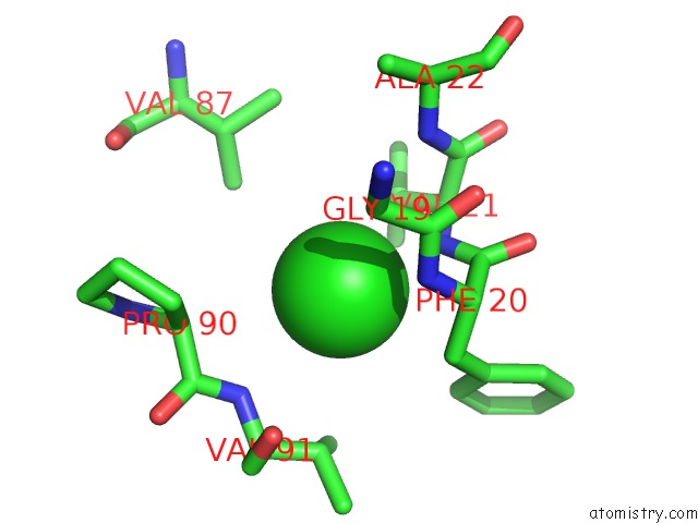
Mono view
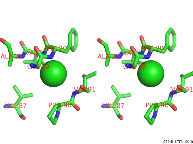
Stereo pair view
Mono view
Stereo pair view
A full contact list of Chlorine with other atoms in the Cl binding
site number 1 of Structure of Apo Anthocyanidin Reductase From Vitis Vinifera within 5.0Å range:
|
Chlorine binding site 2 out of 2 in 3hfs
Go back to
Chlorine binding site 2 out
of 2 in the Structure of Apo Anthocyanidin Reductase From Vitis Vinifera
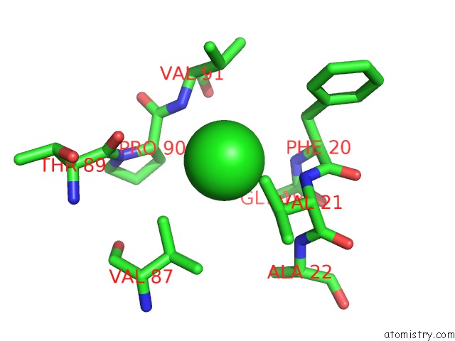
Mono view
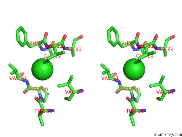
Stereo pair view
Mono view
Stereo pair view
A full contact list of Chlorine with other atoms in the Cl binding
site number 2 of Structure of Apo Anthocyanidin Reductase From Vitis Vinifera within 5.0Å range:
|
Reference:
M.Gargouri,
C.Manigand,
C.Mauge,
T.Granier,
B.Langlois D'estaintot,
O.Cala,
I.Pianet,
K.Bathany,
J.Chaudiere,
B.Gallois.
Structure and Epimerase Activity of Anthocyanidin Reductase From Vitis Vinifera. Acta Crystallogr.,Sect.D V. 65 989 2009.
ISSN: ISSN 0907-4449
PubMed: 19690377
DOI: 10.1107/S0907444909025013
Page generated: Sat Jul 20 20:47:50 2024
ISSN: ISSN 0907-4449
PubMed: 19690377
DOI: 10.1107/S0907444909025013
Last articles
Zn in 9J0NZn in 9J0O
Zn in 9J0P
Zn in 9FJX
Zn in 9EKB
Zn in 9C0F
Zn in 9CAH
Zn in 9CH0
Zn in 9CH3
Zn in 9CH1