Chlorine »
PDB 3hl8-3hwl »
3hmp »
Chlorine in PDB 3hmp: Crystal Structure of Human MPS1 Catalytic Domain in Complex with A Quinazolin Ligand Compound 4
Enzymatic activity of Crystal Structure of Human MPS1 Catalytic Domain in Complex with A Quinazolin Ligand Compound 4
All present enzymatic activity of Crystal Structure of Human MPS1 Catalytic Domain in Complex with A Quinazolin Ligand Compound 4:
2.7.12.1;
2.7.12.1;
Protein crystallography data
The structure of Crystal Structure of Human MPS1 Catalytic Domain in Complex with A Quinazolin Ligand Compound 4, PDB code: 3hmp
was solved by
M.L.H.Chu,
L.M.G.Chavas,
D.H.Williams,
L.Tabernero,
P.A.Eyers,
with X-Ray Crystallography technique. A brief refinement statistics is given in the table below:
| Resolution Low / High (Å) | 60.15 / 2.30 |
| Space group | I 2 2 2 |
| Cell size a, b, c (Å), α, β, γ (°) | 71.370, 103.930, 111.730, 90.00, 90.00, 90.00 |
| R / Rfree (%) | 21 / 24.8 |
Chlorine Binding Sites:
The binding sites of Chlorine atom in the Crystal Structure of Human MPS1 Catalytic Domain in Complex with A Quinazolin Ligand Compound 4
(pdb code 3hmp). This binding sites where shown within
5.0 Angstroms radius around Chlorine atom.
In total only one binding site of Chlorine was determined in the Crystal Structure of Human MPS1 Catalytic Domain in Complex with A Quinazolin Ligand Compound 4, PDB code: 3hmp:
In total only one binding site of Chlorine was determined in the Crystal Structure of Human MPS1 Catalytic Domain in Complex with A Quinazolin Ligand Compound 4, PDB code: 3hmp:
Chlorine binding site 1 out of 1 in 3hmp
Go back to
Chlorine binding site 1 out
of 1 in the Crystal Structure of Human MPS1 Catalytic Domain in Complex with A Quinazolin Ligand Compound 4
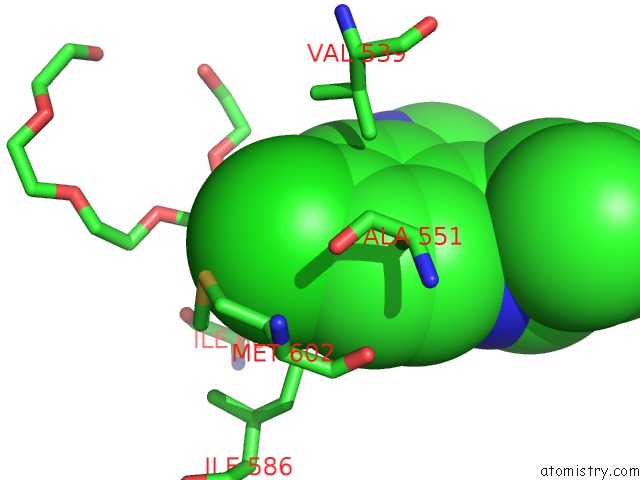
Mono view
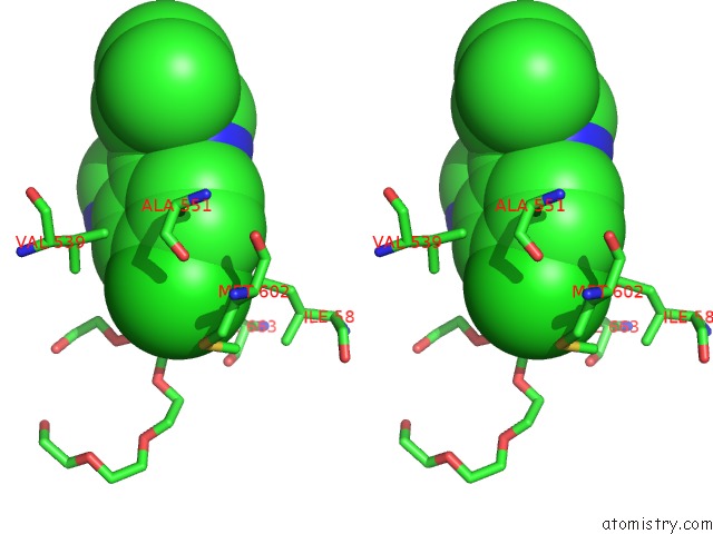
Stereo pair view
Mono view
Stereo pair view
A full contact list of Chlorine with other atoms in the Cl binding
site number 1 of Crystal Structure of Human MPS1 Catalytic Domain in Complex with A Quinazolin Ligand Compound 4 within 5.0Å range:
|
Reference:
M.L.Chu,
Z.Lang,
L.M.Chavas,
J.Neres,
O.S.Fedorova,
L.Tabernero,
M.Cherry,
D.H.Williams,
K.T.Douglas,
P.A.Eyers.
Biophysical and X-Ray Crystallographic Analysis of MPS1 Kinase Inhibitor Complexes. Biochemistry V. 49 1689 2010.
ISSN: ISSN 0006-2960
PubMed: 20099905
DOI: 10.1021/BI901970C
Page generated: Sat Jul 20 20:58:50 2024
ISSN: ISSN 0006-2960
PubMed: 20099905
DOI: 10.1021/BI901970C
Last articles
Zn in 9J0NZn in 9J0O
Zn in 9J0P
Zn in 9FJX
Zn in 9EKB
Zn in 9C0F
Zn in 9CAH
Zn in 9CH0
Zn in 9CH3
Zn in 9CH1