Chlorine »
PDB 3hwp-3i67 »
3i2a »
Chlorine in PDB 3i2a: Crystal Structure of A Chimeric Trypsin Inhibitor Protein Sti(L)- Wci(S)
Protein crystallography data
The structure of Crystal Structure of A Chimeric Trypsin Inhibitor Protein Sti(L)- Wci(S), PDB code: 3i2a
was solved by
U.Sen,
S.Khamrui,
with X-Ray Crystallography technique. A brief refinement statistics is given in the table below:
| Resolution Low / High (Å) | 19.96 / 2.30 |
| Space group | I 4 |
| Cell size a, b, c (Å), α, β, γ (°) | 141.383, 141.383, 46.716, 90.00, 90.00, 90.00 |
| R / Rfree (%) | 23.4 / 28.9 |
Chlorine Binding Sites:
The binding sites of Chlorine atom in the Crystal Structure of A Chimeric Trypsin Inhibitor Protein Sti(L)- Wci(S)
(pdb code 3i2a). This binding sites where shown within
5.0 Angstroms radius around Chlorine atom.
In total 2 binding sites of Chlorine where determined in the Crystal Structure of A Chimeric Trypsin Inhibitor Protein Sti(L)- Wci(S), PDB code: 3i2a:
Jump to Chlorine binding site number: 1; 2;
In total 2 binding sites of Chlorine where determined in the Crystal Structure of A Chimeric Trypsin Inhibitor Protein Sti(L)- Wci(S), PDB code: 3i2a:
Jump to Chlorine binding site number: 1; 2;
Chlorine binding site 1 out of 2 in 3i2a
Go back to
Chlorine binding site 1 out
of 2 in the Crystal Structure of A Chimeric Trypsin Inhibitor Protein Sti(L)- Wci(S)
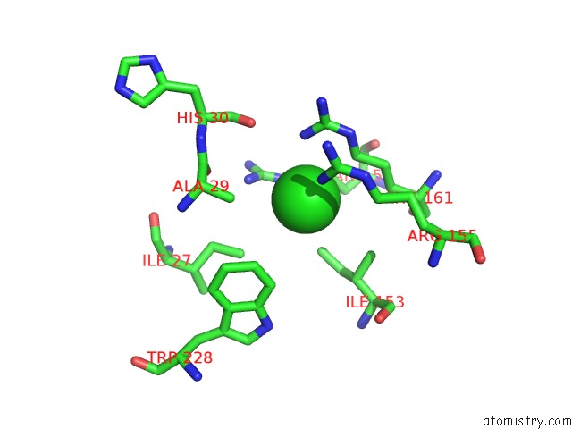
Mono view
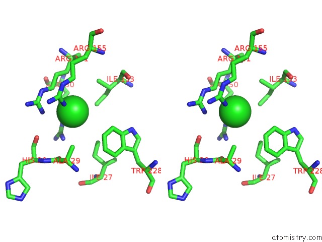
Stereo pair view
Mono view
Stereo pair view
A full contact list of Chlorine with other atoms in the Cl binding
site number 1 of Crystal Structure of A Chimeric Trypsin Inhibitor Protein Sti(L)- Wci(S) within 5.0Å range:
|
Chlorine binding site 2 out of 2 in 3i2a
Go back to
Chlorine binding site 2 out
of 2 in the Crystal Structure of A Chimeric Trypsin Inhibitor Protein Sti(L)- Wci(S)
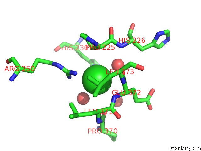
Mono view
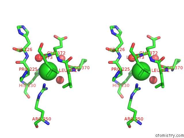
Stereo pair view
Mono view
Stereo pair view
A full contact list of Chlorine with other atoms in the Cl binding
site number 2 of Crystal Structure of A Chimeric Trypsin Inhibitor Protein Sti(L)- Wci(S) within 5.0Å range:
|
Reference:
S.Khamrui,
S.Majumder,
J.Dasgupta,
J.K.Dattagupta,
U.Sen.
Identification of A Novel Set of Scaffolding Residues That Are Instrumental For the Inhibitory Property of Kunitz (Sti) Inhibitors. Protein Sci. V. 19 593 2010.
ISSN: ISSN 0961-8368
PubMed: 20073082
DOI: 10.1002/PRO.338
Page generated: Sat Jul 20 21:11:21 2024
ISSN: ISSN 0961-8368
PubMed: 20073082
DOI: 10.1002/PRO.338
Last articles
Zn in 9J0NZn in 9J0O
Zn in 9J0P
Zn in 9FJX
Zn in 9EKB
Zn in 9C0F
Zn in 9CAH
Zn in 9CH0
Zn in 9CH3
Zn in 9CH1