Chlorine »
PDB 3hwp-3i67 »
3i46 »
Chlorine in PDB 3i46: Crystal Structure of Beta Toxin From Staphylococcus Aureus F277A, P278A Mutant with Bound Calcium Ions
Protein crystallography data
The structure of Crystal Structure of Beta Toxin From Staphylococcus Aureus F277A, P278A Mutant with Bound Calcium Ions, PDB code: 3i46
was solved by
M.Huseby,
K.Shi,
A.C.Kruse,
D.H.Ohlendorf,
with X-Ray Crystallography technique. A brief refinement statistics is given in the table below:
| Resolution Low / High (Å) | 34.04 / 2.60 |
| Space group | P 21 21 21 |
| Cell size a, b, c (Å), α, β, γ (°) | 68.093, 70.195, 129.584, 90.00, 90.00, 90.00 |
| R / Rfree (%) | 22.1 / 27.5 |
Other elements in 3i46:
The structure of Crystal Structure of Beta Toxin From Staphylococcus Aureus F277A, P278A Mutant with Bound Calcium Ions also contains other interesting chemical elements:
| Calcium | (Ca) | 4 atoms |
Chlorine Binding Sites:
The binding sites of Chlorine atom in the Crystal Structure of Beta Toxin From Staphylococcus Aureus F277A, P278A Mutant with Bound Calcium Ions
(pdb code 3i46). This binding sites where shown within
5.0 Angstroms radius around Chlorine atom.
In total 2 binding sites of Chlorine where determined in the Crystal Structure of Beta Toxin From Staphylococcus Aureus F277A, P278A Mutant with Bound Calcium Ions, PDB code: 3i46:
Jump to Chlorine binding site number: 1; 2;
In total 2 binding sites of Chlorine where determined in the Crystal Structure of Beta Toxin From Staphylococcus Aureus F277A, P278A Mutant with Bound Calcium Ions, PDB code: 3i46:
Jump to Chlorine binding site number: 1; 2;
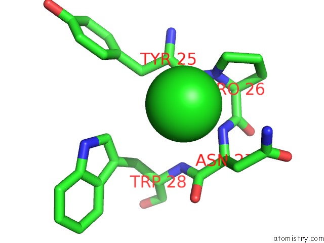
Chlorine binding site 1 out of 2 in 3i46
Go back to
Chlorine binding site 1 out
of 2 in the Crystal Structure of Beta Toxin From Staphylococcus Aureus F277A, P278A Mutant with Bound Calcium Ions
Mono view

Stereo pair view
Mono view
Stereo pair view
A full contact list of Chlorine with other atoms in the Cl binding
site number 1 of Crystal Structure of Beta Toxin From Staphylococcus Aureus F277A, P278A Mutant with Bound Calcium Ions within 5.0Å range:
|
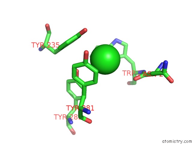
Chlorine binding site 2 out of 2 in 3i46
Go back to
Chlorine binding site 2 out
of 2 in the Crystal Structure of Beta Toxin From Staphylococcus Aureus F277A, P278A Mutant with Bound Calcium Ions
Mono view

Stereo pair view
Mono view
Stereo pair view
A full contact list of Chlorine with other atoms in the Cl binding
site number 2 of Crystal Structure of Beta Toxin From Staphylococcus Aureus F277A, P278A Mutant with Bound Calcium Ions within 5.0Å range:
|
Reference:
M.Huseby,
K.Shi,
A.C.Kruse,
J.Digre,
F.Mengistu,
G.A.Bohach,
P.S.Schlievert,
D.H.Ohlendorf,
C.A.Earhart.
Structure and Biological Functions of Beta Toxin From Staphylococcus Aureus: Role of the Hydrophobic Beta Hairpin in Virulence To Be Published.
Page generated: Sat Jul 20 21:15:33 2024
Last articles
Zn in 9J0NZn in 9J0O
Zn in 9J0P
Zn in 9FJX
Zn in 9EKB
Zn in 9C0F
Zn in 9CAH
Zn in 9CH0
Zn in 9CH3
Zn in 9CH1