Chlorine »
PDB 3i6d-3idv »
3id7 »
Chlorine in PDB 3id7: Crystal Structure of Renal Dipeptidase From Streptomyces Coelicolor A3(2)
Protein crystallography data
The structure of Crystal Structure of Renal Dipeptidase From Streptomyces Coelicolor A3(2), PDB code: 3id7
was solved by
A.A.Fedorov,
E.V.Fedorov,
J.Cummings,
F.M.Raushel,
S.C.Almo,
with X-Ray Crystallography technique. A brief refinement statistics is given in the table below:
| Resolution Low / High (Å) | 24.99 / 1.30 |
| Space group | P 31 2 1 |
| Cell size a, b, c (Å), α, β, γ (°) | 96.747, 96.747, 104.721, 90.00, 90.00, 120.00 |
| R / Rfree (%) | 18.4 / 19.8 |
Other elements in 3id7:
The structure of Crystal Structure of Renal Dipeptidase From Streptomyces Coelicolor A3(2) also contains other interesting chemical elements:
| Zinc | (Zn) | 2 atoms |
Chlorine Binding Sites:
The binding sites of Chlorine atom in the Crystal Structure of Renal Dipeptidase From Streptomyces Coelicolor A3(2)
(pdb code 3id7). This binding sites where shown within
5.0 Angstroms radius around Chlorine atom.
In total only one binding site of Chlorine was determined in the Crystal Structure of Renal Dipeptidase From Streptomyces Coelicolor A3(2), PDB code: 3id7:
In total only one binding site of Chlorine was determined in the Crystal Structure of Renal Dipeptidase From Streptomyces Coelicolor A3(2), PDB code: 3id7:
Chlorine binding site 1 out of 1 in 3id7
Go back to
Chlorine binding site 1 out
of 1 in the Crystal Structure of Renal Dipeptidase From Streptomyces Coelicolor A3(2)
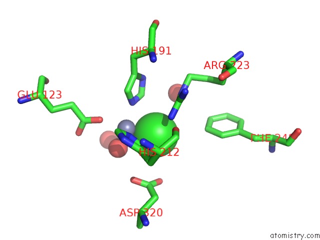
Mono view
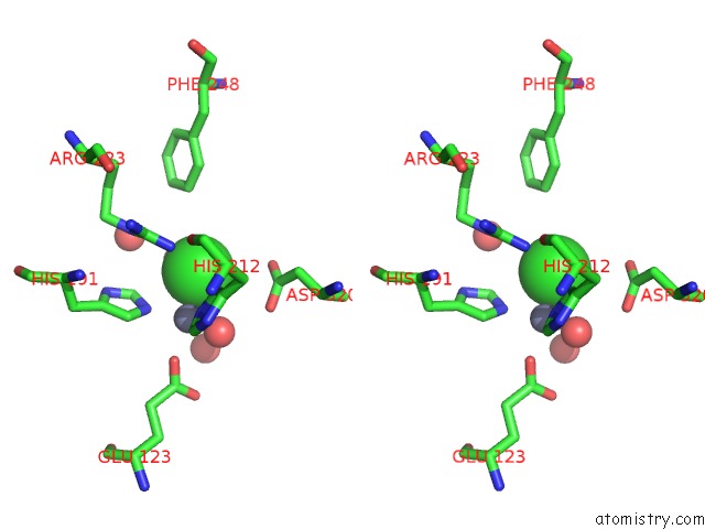
Stereo pair view
Mono view
Stereo pair view
A full contact list of Chlorine with other atoms in the Cl binding
site number 1 of Crystal Structure of Renal Dipeptidase From Streptomyces Coelicolor A3(2) within 5.0Å range:
|
Reference:
J.A.Cummings,
T.T.Nguyen,
A.A.Fedorov,
P.Kolb,
C.Xu,
E.V.Fedorov,
B.K.Shoichet,
D.P.Barondeau,
S.C.Almo,
F.M.Raushel.
Structure, Mechanism, and Substrate Profile For SCO3058: the Closest Bacterial Homologue to Human Renal Dipeptidase . Biochemistry V. 49 611 2010.
ISSN: ISSN 0006-2960
PubMed: 20000809
DOI: 10.1021/BI901935Y
Page generated: Sat Jul 20 21:28:14 2024
ISSN: ISSN 0006-2960
PubMed: 20000809
DOI: 10.1021/BI901935Y
Last articles
Ca in 2RJPCa in 2RO9
Ca in 2RO8
Ca in 2RLD
Ca in 2RNT
Ca in 2RJQ
Ca in 2RIE
Ca in 2RID
Ca in 2RJI
Ca in 2RIC