Chlorine »
PDB 3ie5-3ili »
3ie5 »
Chlorine in PDB 3ie5: Crystal Structure of Hyp-1 Protein From Hypericum Perforatum (St John'S Wort) Involved in Hypericin Biosynthesis
Protein crystallography data
The structure of Crystal Structure of Hyp-1 Protein From Hypericum Perforatum (St John'S Wort) Involved in Hypericin Biosynthesis, PDB code: 3ie5
was solved by
K.Michalska,
H.Fernandes,
M.M.Sikorski,
M.Jaskolski,
with X-Ray Crystallography technique. A brief refinement statistics is given in the table below:
| Resolution Low / High (Å) | 27.66 / 1.69 |
| Space group | P 21 21 21 |
| Cell size a, b, c (Å), α, β, γ (°) | 37.538, 76.713, 119.799, 90.00, 90.00, 90.00 |
| R / Rfree (%) | 17 / 20.6 |
Chlorine Binding Sites:
The binding sites of Chlorine atom in the Crystal Structure of Hyp-1 Protein From Hypericum Perforatum (St John'S Wort) Involved in Hypericin Biosynthesis
(pdb code 3ie5). This binding sites where shown within
5.0 Angstroms radius around Chlorine atom.
In total only one binding site of Chlorine was determined in the Crystal Structure of Hyp-1 Protein From Hypericum Perforatum (St John'S Wort) Involved in Hypericin Biosynthesis, PDB code: 3ie5:
In total only one binding site of Chlorine was determined in the Crystal Structure of Hyp-1 Protein From Hypericum Perforatum (St John'S Wort) Involved in Hypericin Biosynthesis, PDB code: 3ie5:
Chlorine binding site 1 out of 1 in 3ie5
Go back to
Chlorine binding site 1 out
of 1 in the Crystal Structure of Hyp-1 Protein From Hypericum Perforatum (St John'S Wort) Involved in Hypericin Biosynthesis
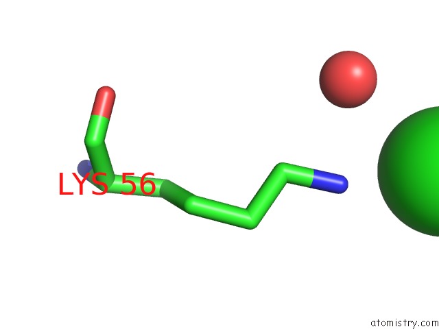
Mono view
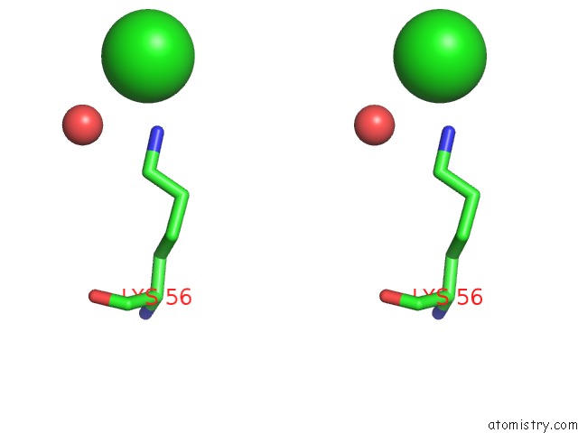
Stereo pair view
Mono view
Stereo pair view
A full contact list of Chlorine with other atoms in the Cl binding
site number 1 of Crystal Structure of Hyp-1 Protein From Hypericum Perforatum (St John'S Wort) Involved in Hypericin Biosynthesis within 5.0Å range:
|
Reference:
K.Michalska,
H.Fernandes,
M.M.Sikorski,
M.Jaskolski.
Crystal Structure of Hyp-1, A St. John'S Wort Protein Implicated in the Biosynthesis of Hypericin J.Struct.Biol. V. 169 161 2010.
ISSN: ISSN 1047-8477
PubMed: 19853038
DOI: 10.1016/J.JSB.2009.10.008
Page generated: Sat Jul 20 21:29:53 2024
ISSN: ISSN 1047-8477
PubMed: 19853038
DOI: 10.1016/J.JSB.2009.10.008
Last articles
Zn in 9J0NZn in 9J0O
Zn in 9J0P
Zn in 9FJX
Zn in 9EKB
Zn in 9C0F
Zn in 9CAH
Zn in 9CH0
Zn in 9CH3
Zn in 9CH1