Chlorine »
PDB 3ilj-3isv »
3irt »
Chlorine in PDB 3irt: Crystal Structure of the I93M Mutant of Ubiquitin Carboxy-Terminal Hydrolase L1
Enzymatic activity of Crystal Structure of the I93M Mutant of Ubiquitin Carboxy-Terminal Hydrolase L1
All present enzymatic activity of Crystal Structure of the I93M Mutant of Ubiquitin Carboxy-Terminal Hydrolase L1:
3.4.19.12;
3.4.19.12;
Protein crystallography data
The structure of Crystal Structure of the I93M Mutant of Ubiquitin Carboxy-Terminal Hydrolase L1, PDB code: 3irt
was solved by
C.W.Davies,
T.K.Maiti,
C.Das,
with X-Ray Crystallography technique. A brief refinement statistics is given in the table below:
| Resolution Low / High (Å) | 41.75 / 2.80 |
| Space group | P 4 21 2 |
| Cell size a, b, c (Å), α, β, γ (°) | 109.938, 109.938, 79.040, 90.00, 90.00, 90.00 |
| R / Rfree (%) | 21.1 / 25.4 |
Chlorine Binding Sites:
The binding sites of Chlorine atom in the Crystal Structure of the I93M Mutant of Ubiquitin Carboxy-Terminal Hydrolase L1
(pdb code 3irt). This binding sites where shown within
5.0 Angstroms radius around Chlorine atom.
In total 2 binding sites of Chlorine where determined in the Crystal Structure of the I93M Mutant of Ubiquitin Carboxy-Terminal Hydrolase L1, PDB code: 3irt:
Jump to Chlorine binding site number: 1; 2;
In total 2 binding sites of Chlorine where determined in the Crystal Structure of the I93M Mutant of Ubiquitin Carboxy-Terminal Hydrolase L1, PDB code: 3irt:
Jump to Chlorine binding site number: 1; 2;
Chlorine binding site 1 out of 2 in 3irt
Go back to
Chlorine binding site 1 out
of 2 in the Crystal Structure of the I93M Mutant of Ubiquitin Carboxy-Terminal Hydrolase L1
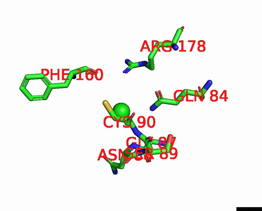
Mono view
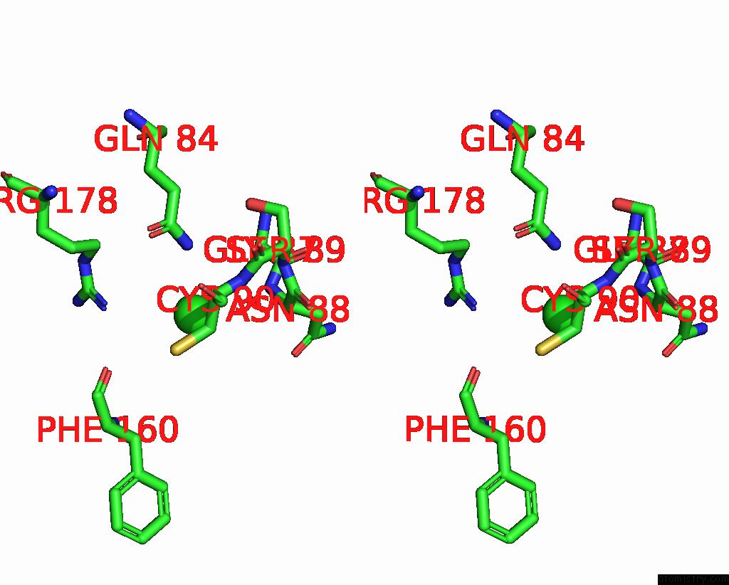
Stereo pair view
Mono view
Stereo pair view
A full contact list of Chlorine with other atoms in the Cl binding
site number 1 of Crystal Structure of the I93M Mutant of Ubiquitin Carboxy-Terminal Hydrolase L1 within 5.0Å range:
|
Chlorine binding site 2 out of 2 in 3irt
Go back to
Chlorine binding site 2 out
of 2 in the Crystal Structure of the I93M Mutant of Ubiquitin Carboxy-Terminal Hydrolase L1
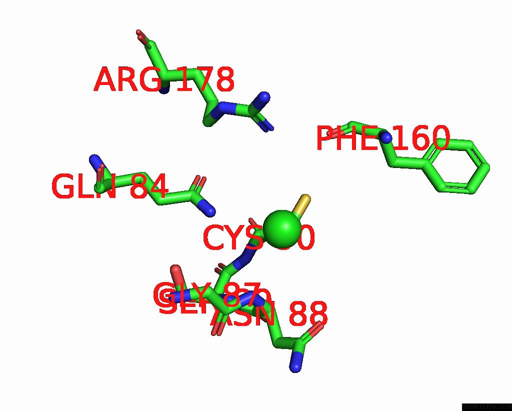
Mono view
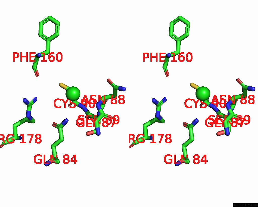
Stereo pair view
Mono view
Stereo pair view
A full contact list of Chlorine with other atoms in the Cl binding
site number 2 of Crystal Structure of the I93M Mutant of Ubiquitin Carboxy-Terminal Hydrolase L1 within 5.0Å range:
|
Reference:
D.A.Boudreaux,
T.K.Maiti,
C.W.Davies,
C.Das.
Ubiquitin Vinyl Methyl Ester Binding Orients the Misaligned Active Site of the Ubiquitin Hydrolase UCHL1 Into Productive Conformation. Proc.Natl.Acad.Sci.Usa V. 107 9117 2010.
ISSN: ISSN 0027-8424
PubMed: 20439756
DOI: 10.1073/PNAS.0910870107
Page generated: Sat Jul 20 21:51:00 2024
ISSN: ISSN 0027-8424
PubMed: 20439756
DOI: 10.1073/PNAS.0910870107
Last articles
Zn in 9J0NZn in 9J0O
Zn in 9J0P
Zn in 9FJX
Zn in 9EKB
Zn in 9C0F
Zn in 9CAH
Zn in 9CH0
Zn in 9CH3
Zn in 9CH1