Chlorine »
PDB 3jq0-3k1s »
3jqy »
Chlorine in PDB 3jqy: Crystal Strucutre of the Polysia Specific Acetyltransferase Neuo
Enzymatic activity of Crystal Strucutre of the Polysia Specific Acetyltransferase Neuo
All present enzymatic activity of Crystal Strucutre of the Polysia Specific Acetyltransferase Neuo:
2.3.1.136;
2.3.1.136;
Protein crystallography data
The structure of Crystal Strucutre of the Polysia Specific Acetyltransferase Neuo, PDB code: 3jqy
was solved by
E.-C.Schulz,
A.Bergfeld,
M.Muehlenhoff,
R.Ficner,
with X-Ray Crystallography technique. A brief refinement statistics is given in the table below:
| Resolution Low / High (Å) | 37.36 / 1.70 |
| Space group | P 1 21 1 |
| Cell size a, b, c (Å), α, β, γ (°) | 62.833, 88.366, 72.996, 90.00, 106.62, 90.00 |
| R / Rfree (%) | 16.6 / 19.5 |
Chlorine Binding Sites:
The binding sites of Chlorine atom in the Crystal Strucutre of the Polysia Specific Acetyltransferase Neuo
(pdb code 3jqy). This binding sites where shown within
5.0 Angstroms radius around Chlorine atom.
In total 2 binding sites of Chlorine where determined in the Crystal Strucutre of the Polysia Specific Acetyltransferase Neuo, PDB code: 3jqy:
Jump to Chlorine binding site number: 1; 2;
In total 2 binding sites of Chlorine where determined in the Crystal Strucutre of the Polysia Specific Acetyltransferase Neuo, PDB code: 3jqy:
Jump to Chlorine binding site number: 1; 2;
Chlorine binding site 1 out of 2 in 3jqy
Go back to
Chlorine binding site 1 out
of 2 in the Crystal Strucutre of the Polysia Specific Acetyltransferase Neuo
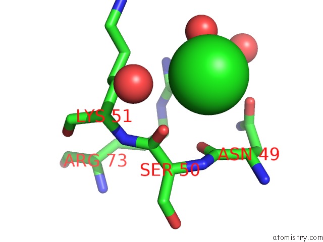
Mono view
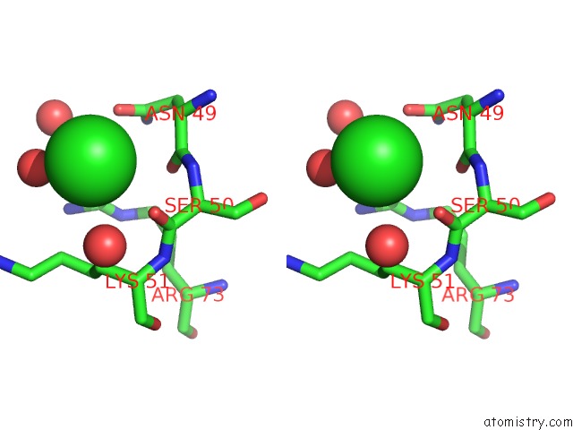
Stereo pair view
Mono view
Stereo pair view
A full contact list of Chlorine with other atoms in the Cl binding
site number 1 of Crystal Strucutre of the Polysia Specific Acetyltransferase Neuo within 5.0Å range:
|
Chlorine binding site 2 out of 2 in 3jqy
Go back to
Chlorine binding site 2 out
of 2 in the Crystal Strucutre of the Polysia Specific Acetyltransferase Neuo
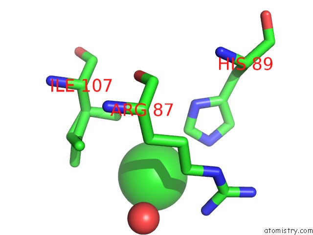
Mono view
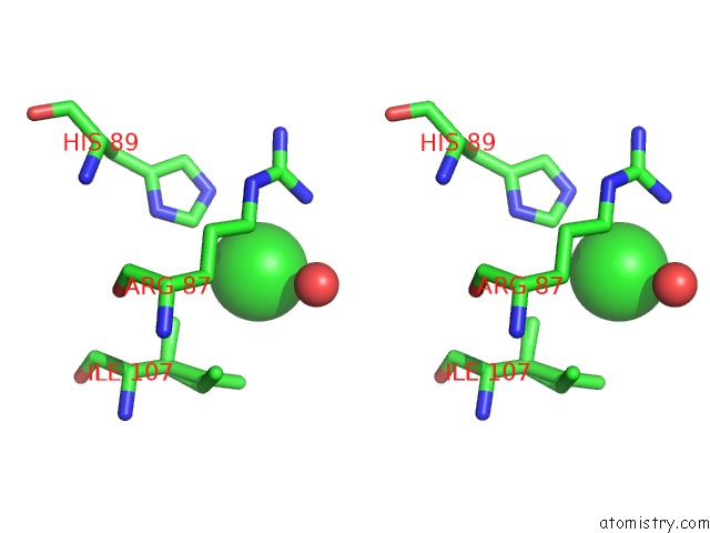
Stereo pair view
Mono view
Stereo pair view
A full contact list of Chlorine with other atoms in the Cl binding
site number 2 of Crystal Strucutre of the Polysia Specific Acetyltransferase Neuo within 5.0Å range:
|
Reference:
E.C.Schulz,
A.K.Bergfeld,
R.Ficner,
M.Muhlenhoff.
Crystal Structure Analysis of the Polysialic Acid Specific O-Acetyltransferase Neuo Plos One V. 6 17403 2011.
ISSN: ESSN 1932-6203
PubMed: 21390252
DOI: 10.1371/JOURNAL.PONE.0017403
Page generated: Fri Jul 11 06:43:39 2025
ISSN: ESSN 1932-6203
PubMed: 21390252
DOI: 10.1371/JOURNAL.PONE.0017403
Last articles
Fe in 2YXOFe in 2YRS
Fe in 2YXC
Fe in 2YNM
Fe in 2YVJ
Fe in 2YP1
Fe in 2YU2
Fe in 2YU1
Fe in 2YQB
Fe in 2YOO