Chlorine »
PDB 3jq0-3k1s »
3jv9 »
Chlorine in PDB 3jv9: The Structure of A Reduced Form of Oxyr From N. Meningitidis
Protein crystallography data
The structure of The Structure of A Reduced Form of Oxyr From N. Meningitidis, PDB code: 3jv9
was solved by
S.Sainsbury,
J.Ren,
D.I.Stuart,
R.J.Owens,
Oxford Protein Productionfacility (Oppf),
with X-Ray Crystallography technique. A brief refinement statistics is given in the table below:
| Resolution Low / High (Å) | 29.80 / 2.39 |
| Space group | P 1 21 1 |
| Cell size a, b, c (Å), α, β, γ (°) | 49.813, 56.075, 81.247, 90.00, 104.91, 90.00 |
| R / Rfree (%) | 22.4 / 28.6 |
Chlorine Binding Sites:
The binding sites of Chlorine atom in the The Structure of A Reduced Form of Oxyr From N. Meningitidis
(pdb code 3jv9). This binding sites where shown within
5.0 Angstroms radius around Chlorine atom.
In total 2 binding sites of Chlorine where determined in the The Structure of A Reduced Form of Oxyr From N. Meningitidis, PDB code: 3jv9:
Jump to Chlorine binding site number: 1; 2;
In total 2 binding sites of Chlorine where determined in the The Structure of A Reduced Form of Oxyr From N. Meningitidis, PDB code: 3jv9:
Jump to Chlorine binding site number: 1; 2;
Chlorine binding site 1 out of 2 in 3jv9
Go back to
Chlorine binding site 1 out
of 2 in the The Structure of A Reduced Form of Oxyr From N. Meningitidis
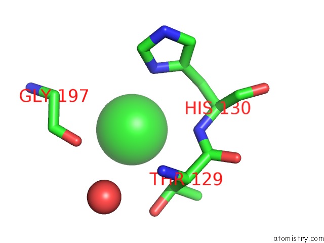
Mono view
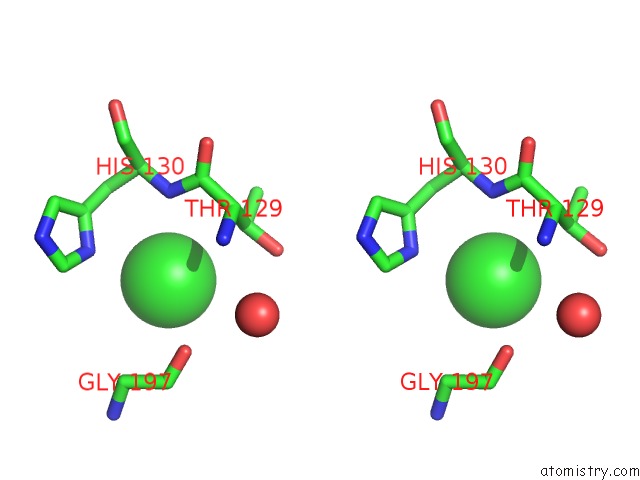
Stereo pair view
Mono view
Stereo pair view
A full contact list of Chlorine with other atoms in the Cl binding
site number 1 of The Structure of A Reduced Form of Oxyr From N. Meningitidis within 5.0Å range:
|
Chlorine binding site 2 out of 2 in 3jv9
Go back to
Chlorine binding site 2 out
of 2 in the The Structure of A Reduced Form of Oxyr From N. Meningitidis
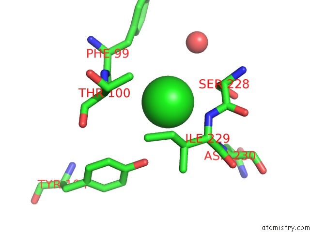
Mono view
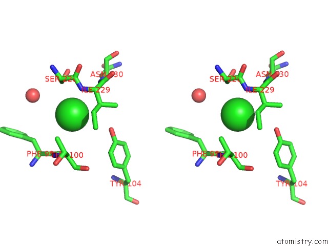
Stereo pair view
Mono view
Stereo pair view
A full contact list of Chlorine with other atoms in the Cl binding
site number 2 of The Structure of A Reduced Form of Oxyr From N. Meningitidis within 5.0Å range:
|
Reference:
S.Sainsbury,
J.Ren,
J.E.Nettleship,
N.J.Saunders,
D.I.Stuart,
R.J.Owens.
The Structure of A Reduced Form of Oxyr From Neisseria Meningitidis Bmc Struct.Biol. V. 10 10 2010.
ISSN: ESSN 1472-6807
PubMed: 20478059
DOI: 10.1186/1472-6807-10-10
Page generated: Sat Jul 20 22:13:43 2024
ISSN: ESSN 1472-6807
PubMed: 20478059
DOI: 10.1186/1472-6807-10-10
Last articles
Zn in 9J0NZn in 9J0O
Zn in 9J0P
Zn in 9FJX
Zn in 9EKB
Zn in 9C0F
Zn in 9CAH
Zn in 9CH0
Zn in 9CH3
Zn in 9CH1