Chlorine »
PDB 3k1t-3k9t »
3k2o »
Chlorine in PDB 3k2o: Structure of An Oxygenase
Protein crystallography data
The structure of Structure of An Oxygenase, PDB code: 3k2o
was solved by
T.Krojer,
M.A.Mcdonough,
I.J.Clifton,
M.Mantri,
S.S.Ng,
A.C.W.Pike,
D.S.Butler,
C.J.Webby,
G.Kochan,
C.Bhatia,
J.E.Bray,
A.Chaikuad,
O.Gileadi,
F.Von Delft,
J.Weigelt,
C.H.Arrowsmith,
C.Bountra,
A.M.Edwards,
C.J.Schofield,
K.L.Kavanagh,
U.Oppermann,
with X-Ray Crystallography technique. A brief refinement statistics is given in the table below:
| Resolution Low / High (Å) | 19.85 / 1.75 |
| Space group | P 1 21 1 |
| Cell size a, b, c (Å), α, β, γ (°) | 49.440, 102.030, 98.218, 90.00, 95.89, 90.00 |
| R / Rfree (%) | 19.2 / 22.5 |
Other elements in 3k2o:
The structure of Structure of An Oxygenase also contains other interesting chemical elements:
| Nickel | (Ni) | 2 atoms |
| Sodium | (Na) | 2 atoms |
Chlorine Binding Sites:
The binding sites of Chlorine atom in the Structure of An Oxygenase
(pdb code 3k2o). This binding sites where shown within
5.0 Angstroms radius around Chlorine atom.
In total 6 binding sites of Chlorine where determined in the Structure of An Oxygenase, PDB code: 3k2o:
Jump to Chlorine binding site number: 1; 2; 3; 4; 5; 6;
In total 6 binding sites of Chlorine where determined in the Structure of An Oxygenase, PDB code: 3k2o:
Jump to Chlorine binding site number: 1; 2; 3; 4; 5; 6;
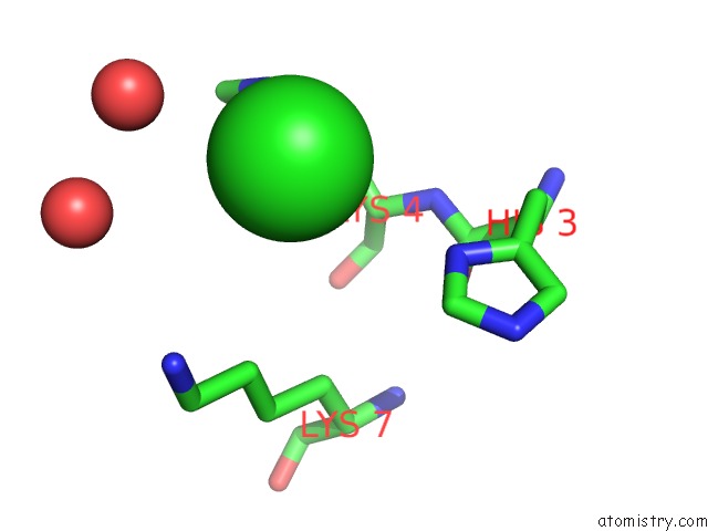
Chlorine binding site 1 out of 6 in 3k2o
Go back to
Chlorine binding site 1 out
of 6 in the Structure of An Oxygenase

Mono view

Stereo pair view
Mono view
Stereo pair view
A full contact list of Chlorine with other atoms in the Cl binding
site number 1 of Structure of An Oxygenase within 5.0Å range:
|
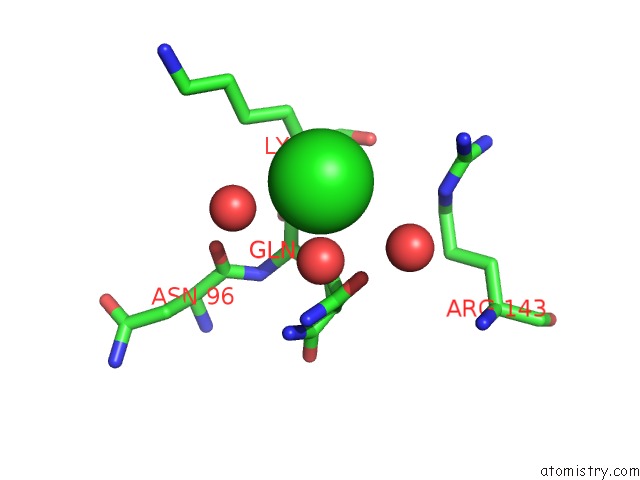
Chlorine binding site 2 out of 6 in 3k2o
Go back to
Chlorine binding site 2 out
of 6 in the Structure of An Oxygenase
Mono view

Stereo pair view
Mono view
Stereo pair view
A full contact list of Chlorine with other atoms in the Cl binding
site number 2 of Structure of An Oxygenase within 5.0Å range:
|
Chlorine binding site 3 out of 6 in 3k2o
Go back to
Chlorine binding site 3 out
of 6 in the Structure of An Oxygenase

Mono view
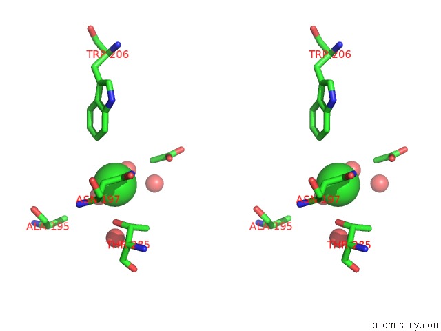
Stereo pair view
Mono view
Stereo pair view
A full contact list of Chlorine with other atoms in the Cl binding
site number 3 of Structure of An Oxygenase within 5.0Å range:
|
Chlorine binding site 4 out of 6 in 3k2o
Go back to
Chlorine binding site 4 out
of 6 in the Structure of An Oxygenase

Mono view

Stereo pair view
Mono view
Stereo pair view
A full contact list of Chlorine with other atoms in the Cl binding
site number 4 of Structure of An Oxygenase within 5.0Å range:
|
Chlorine binding site 5 out of 6 in 3k2o
Go back to
Chlorine binding site 5 out
of 6 in the Structure of An Oxygenase

Mono view
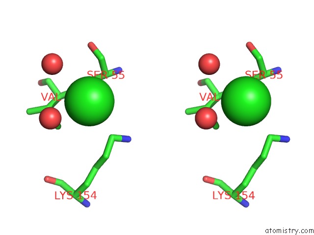
Stereo pair view
Mono view
Stereo pair view
A full contact list of Chlorine with other atoms in the Cl binding
site number 5 of Structure of An Oxygenase within 5.0Å range:
|
Chlorine binding site 6 out of 6 in 3k2o
Go back to
Chlorine binding site 6 out
of 6 in the Structure of An Oxygenase
Mono view

Stereo pair view
Mono view
Stereo pair view
A full contact list of Chlorine with other atoms in the Cl binding
site number 6 of Structure of An Oxygenase within 5.0Å range:
|
Reference:
M.Mantri,
T.Krojer,
E.A.Bagg,
C.A.Webby,
D.S.Butler,
G.Kochan,
K.L.Kavanagh,
U.Oppermann,
M.A.Mcdonough,
C.J.Schofield.
Crystal Structure of the 2-Oxoglutarate- and Fe(II)-Dependent Lysyl Hydroxylase JMJD6. J.Mol.Biol. V. 401 211 2010.
ISSN: ISSN 0022-2836
PubMed: 20685276
DOI: 10.1016/J.JMB.2010.05.054
Page generated: Sat Jul 20 22:23:02 2024
ISSN: ISSN 0022-2836
PubMed: 20685276
DOI: 10.1016/J.JMB.2010.05.054
Last articles
Zn in 9J0NZn in 9J0O
Zn in 9J0P
Zn in 9FJX
Zn in 9EKB
Zn in 9C0F
Zn in 9CAH
Zn in 9CH0
Zn in 9CH3
Zn in 9CH1